

Immunohistochemistry of the lymphoid tissues of the tammar wallaby, Macropus eugenii
- PMID: 12363276
- PMCID: PMC1570915
- DOI: 10.1046/j.1469-7580.2002.00090.x
Immunohistochemistry of the lymphoid tissues of the tammar wallaby, Macropus eugenii
Abstract
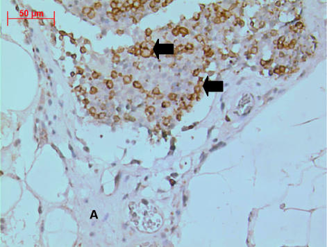
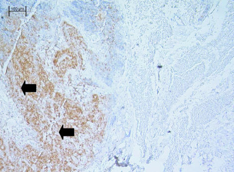
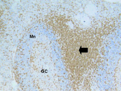
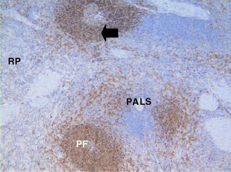
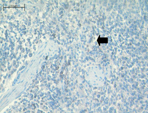
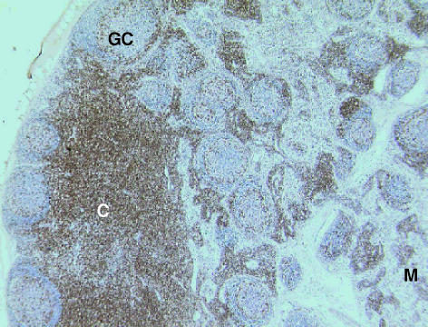
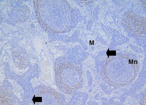
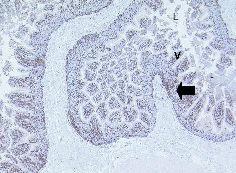
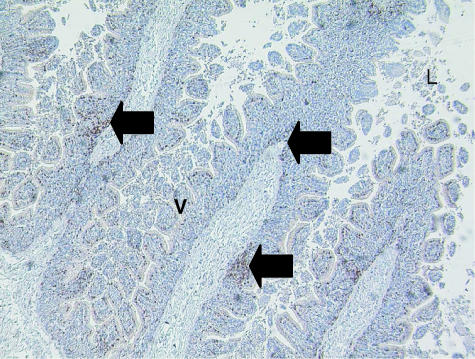
The lymphoid tissues of the metatherian mammal, the adult tammar wallaby, Macropus eugenii, were investigated using immunohistochemical techniques. Five cross-reactive antibodies previously shown to recognize surface markers in marsupial tissues and five previously untested antibodies were used. The distribution of T-cells in the tissue beds of spleen, lymph node, thymus, gut-associated lymphoid tissue (GALT) and bronchus-associated lymphoid tissue (BALT) was documented using antibodies to CD3 and CD5. Similarly, B-cells were identified in the same tissues using anti-CD79b. Antibodies to CD8, CD31, CD79a and CD68 failed to recognize cells in these tissue beds. In general the pattern of cellular distribution identified using these antibodies was similar to that observed in other marsupial and eutherian lymphoid tissues. This study provides further information on the commonality of lymphoid tissue structure in the two major groups of extant mammals, metatherians and eutherians.
Figures
Similar articles
-
The detection of mature T- and B-cells during development of the lymphoid tissues of the tammar wallaby (Macropus eugenii).J Anat. 2003 Jul;203(1):123-31. doi: 10.1046/j.1469-7580.2003.00207.x. J Anat. 2003. PMID: 12892411 Free PMC article.
-
Immunohistochemical localization of T-lymphocyte subsets in the developing lymphoid tissues of the tammar wallaby (Macropus eugenii).Dev Comp Immunol. 2012 Dec;38(4):475-86. doi: 10.1016/j.dci.2012.06.015. Epub 2012 Aug 26. Dev Comp Immunol. 2012. PMID: 22929957
-
Histology and immunohistochemistry of the gut-associated lymphoid tissue of the eastern grey kangaroo, Macropus giganteus.J Anat. 2001 Dec;199(Pt 6):657-62. doi: 10.1046/j.1469-7580.2001.19960657.x. J Anat. 2001. PMID: 11787819 Free PMC article.
-
The development of the immune tissues in marsupial pouch young.J Morphol. 2014 Jul;275(7):822-39. doi: 10.1002/jmor.20250. Epub 2014 Jan 28. J Morphol. 2014. PMID: 24469962 Review.
-
The roles of histology and immunohistology in the investigation of marsupial disease and normal lymphoid tissue.Dev Comp Immunol. 2000 Jul;24(5):455-71. doi: 10.1016/s0145-305x(00)00009-4. Dev Comp Immunol. 2000. PMID: 10785271 Review.
Cited by
-
Transcriptomic analysis supports similar functional roles for the two thymuses of the tammar wallaby.BMC Genomics. 2011 Aug 19;12:420. doi: 10.1186/1471-2164-12-420. BMC Genomics. 2011. PMID: 21854594 Free PMC article.
-
Cathelicidin antimicrobial peptides mediate immune protection in marsupial neonates.Sci Adv. 2025 Apr 18;11(16):eads6359. doi: 10.1126/sciadv.ads6359. Epub 2025 Apr 16. Sci Adv. 2025. PMID: 40238884 Free PMC article.
-
Immunohistochemical Characterization of Immune System Cells in Lymphoid Organs from Roe and Fallow Deer.Animals (Basel). 2022 Nov 7;12(21):3064. doi: 10.3390/ani12213064. Animals (Basel). 2022. PMID: 36359187 Free PMC article.
-
The immune tissues of the endangered red-tailed phascogale (Phascogale calura).J Anat. 2006 Mar;208(3):381-7. doi: 10.1111/j.1469-7580.2006.00530.x. J Anat. 2006. PMID: 16533320 Free PMC article.
-
The appearance and distribution of mature T and B cells in the developing immune tissues of the stripe-faced dunnart (Sminthopsis macroura).J Anat. 2004 Jul;205(1):25-33. doi: 10.1111/j.0021-8782.2004.00310.x. J Anat. 2004. PMID: 15255959 Free PMC article.
References
-
- Adamski FM, Demmer J. Immunological protection of the vulnerable marsupial pouch young: two periods of immune transfer during lactation in the marsupial, Trichosurus vulpecula. Dev. Comparative Immunol. 2000;24:491–502. - PubMed
-
- Armstrong JR, Ferguson MWJ. Ontological wound-healing studies in Monodelphis domestica from birth to adulthood. In: Saunders NR, Hinds L, editors. Marsupial Biology: Recent Research, New Perspectives. Sydney: UNSW Press; 1997. pp. 254–261.
-
- Baker ML, Gemmell E, Gemmell RT. Ontogeny of the brushtail possum, Trichosurus vulpecula. Anat. Record. 1999;256:354–365. - PubMed
-
- Bancroft JD, Stevens AS. Theory and Practice of Histological Techniques. 2. New York: Churchill Livingstone; 1982.
-
- Basden K, Cooper DW, Deane EM. Development of the blood-forming tissues of the tammar wallaby Macropus eugenii. Reprod. Fertil. Dev. 1996;8:989–994. - PubMed
MeSH terms
Substances
LinkOut - more resources
Full Text Sources
Research Materials
Miscellaneous
