. 2002 Dec 6;277(49):47917-27.
doi: 10.1074/jbc.M208191200. Epub 2002 Oct 2.

The Ccz1-Mon1 protein complex is required for the late step of multiple vacuole delivery pathways

Affiliations

The Ccz1-Mon1 protein complex is required for the late step of multiple vacuole delivery pathways

Chao-Wen Wang et al. J Biol Chem. .

Abstract

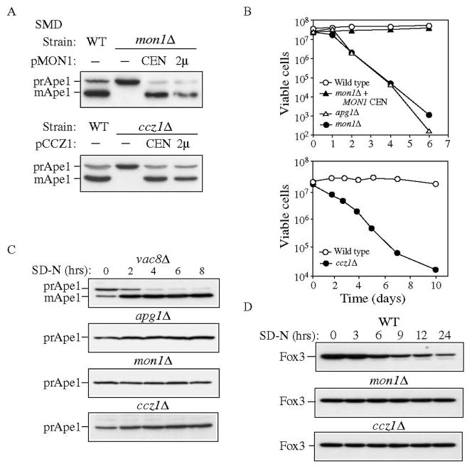
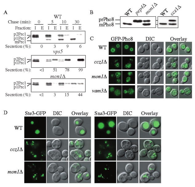
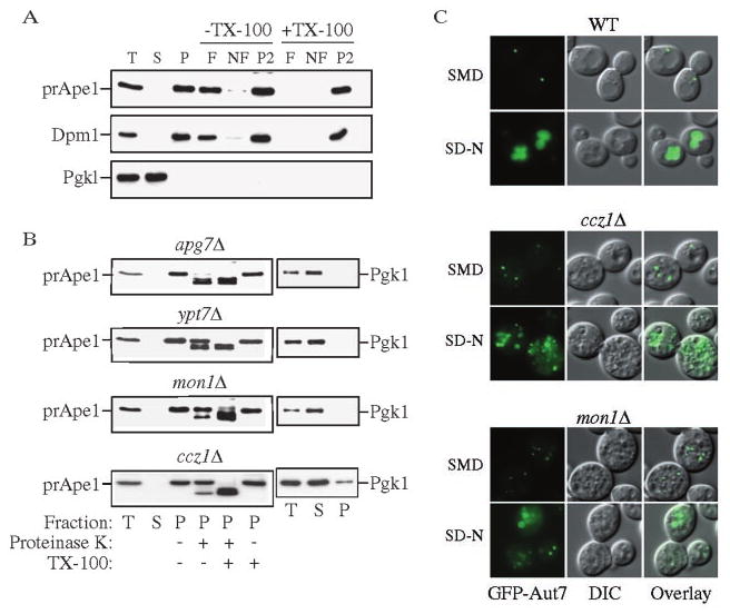
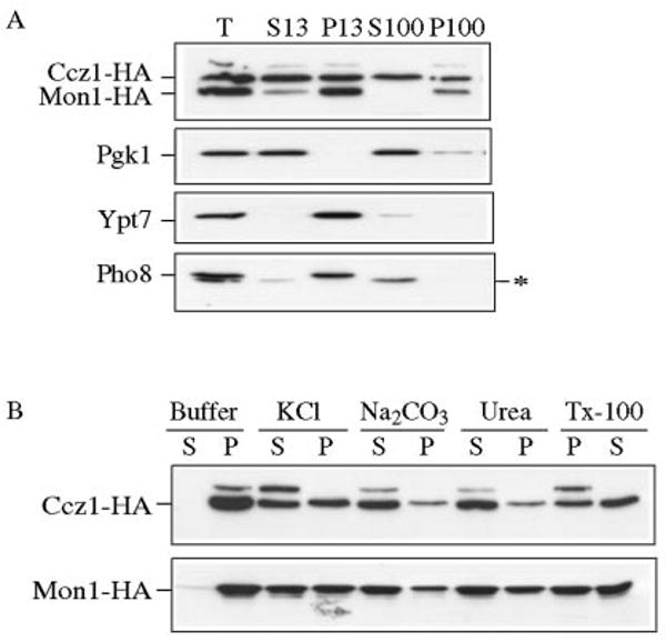
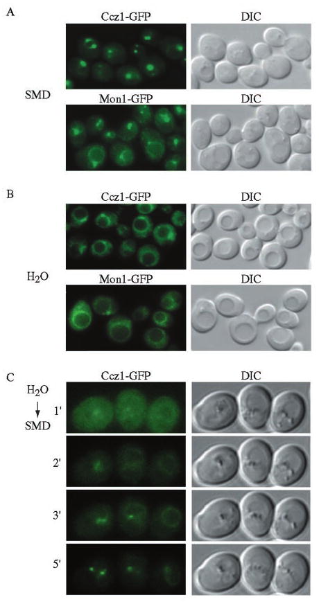
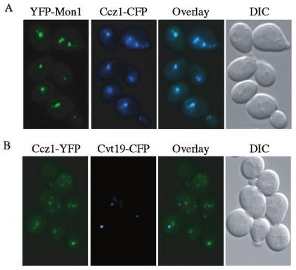
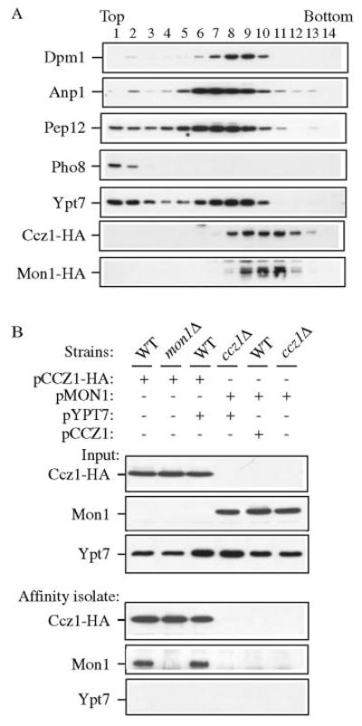
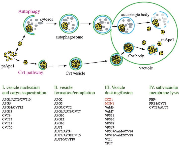
Mon1 and Ccz1 were identified from a gene deletion library as mutants defective in the vacuolar import of aminopeptidase I (Ape1) via the cytoplasm to vacuole targeting (Cvt) pathway. The mon1Delta and ccz1Delta strains also displayed defects in autophagy and pexophagy, degradative pathways that share protein machinery and mechanistic features with the biosynthetic Cvt pathway. Further analyses indicated that Mon1, like Ccz1, was required in nearly all membrane-trafficking pathways where the vacuole represented the terminal acceptor compartment. Accordingly, both deletion strains had kinetic defects in the biosynthetic delivery of resident vacuolar hydrolases through the CPY, ALP, and MVB pathways. Biochemical and microscopy studies suggested that Mon1 and Ccz1 functioned after transport vesicle formation but before (or at) the fusion step with the vacuole. Thus, ccz1Delta and mon1Delta are the first mutants identified in screens for the Cvt and Apg pathways that accumulate precursor Ape1 within completed cytosolic vesicles. Subcellular fractionation and co-immunoprecipitation experiments confirm that Mon1 and Ccz1 physically interact as a stable protein complex termed the Ccz1-Mon1 complex. Microscopy of Ccz1 and Mon1 tagged with a fluorescent marker indicated that the Ccz1-Mon1 complex peripherally associated with a perivacuolar compartment and may attach to the vacuole membrane in agreement with their proposed function in fusion.


Figures

FIG. 1
FIG. 1. The ccz1Δ and mon1Δ strains are defective in the Cvt, autophagy, and pexophagy pathways
A, cloning and characterization of CCZ1 and MON1. Wild type (WT, SEY6210), ccz1Δ (CWY3), and mon1Δ (JSY1) strains and the knockout strains expressing the respective single copy (CEN) or multicopy (2μ) plasmids were grown in SMD medium and analyzed by immunoblot against Ape1. B, mon1Δ and ccz1Δ strain are sensitive to nitrogen-starvation conditions. The wild type, apg1Δ, and mon1Δ strains and the mon1Δ strain harboring pMON1(416) or the wild type and ccz1Δ strains were grown to mid-log phase in SMD medium and shifted to SD-N medium. At the indicated time, aliquots were removed and spread onto YPD plates in triplicate. The number of viable colonies was counted after 2 days incubation at 30 °C. C, mon1Δ and ccz1Δ mutants do not bypass the prApe1 accumulation defect when autophagy is induced. The vac8Δ (D3Y102), apg1Δ (NNY20), ccz1Δ, and mon1Δ strains were grown to mid-log phase in SMD and shifted to SD-N medium. At the indicated time, aliquots were removed and subjected to immunoblot against Ape1. D, mon1Δ and ccz1Δ strains are defective for pexophagy. The wild type, ccz1Δ and mon1Δ strains in the BY4742 background were grown in YPD to mid-log phase, transferred to oleic acid medium to induce peroxisome production and shifted to SD-N. Aliquots were removed at the indicated times and analyzed by Western blot with antiserum to Fox3.
FIG. 2
FIG. 2. Multiple vacuole transport pathways are blocked in the ccz1Δ and mon1Δ strains
A, the mon1Δ strain missorts Prc1 into the extracellular fraction. The wild type (WT, SEY6210), vps5, and mon1Δ (JSY1) strains were grown to mid-log phase and converted into spheroplasts. The spheroplasts were labeled for 5 min and subjected to a non-radioactive chase for the time indicated at 30 °C. Samples were separated into intracellular (I) and extracellular (E) fractions, immunoprecipitated with antiserum to Prc1, and separated by SDS-PAGE. B, mon1Δ and ccz1Δ strains accumulate precursor Pho8. The wild type, pep4Δ (TVY1), ccz1Δ (CWY3), and mon1Δ strains were grown to mid-log phase in SMD medium and analyzed by immunoblot using antiserum against Pho8. C, GFP-Pho8 reaches the vacuoles of the ccz1Δ and mon1Δ strains. Wild type, ccz1Δ, mon1Δ, and vam3Δ (CWY40) strains were transformed with pGFP-Pho8 (426) and grown in SMD medium to mid-log phase followed by fluorescence microscopy. D, endocytic and MVB vesicles accumulated in the ccz1Δ and mon1Δ cells outside of their vacuoles. The wild type, ccz1Δ, and mon1Δ strains expressing the endocytosis pathway marker Ste3-GFP (316), or the MVB pathway marker Sna3 GFP(416) were grown to mid-log phase followed by fluorescence microscopy. DIC, differential interference contrast.
FIG. 3
FIG. 3. Mon1 and Ccz1 are required after completion of Cvt vesicles
A, precursor Ape1 is membrane associated in the mon1Δ strain. The mon1Δ (JSY1) strain was grown to mid-log phase and converted into spheroplasts. The spheroplasts were lysed osmotically and centrifuged through a Ficoll step gradient with or without Triton X-100 as described under “Experimental Procedures.” Membrane-containing float (F), nonfloat (NF), and pellet (P2) fractions were collected and subjected to immunoblot using antisera or antibodies to Ape1, Dpm1, and Pgk1. B, precursor Ape1 is protease-protected in the mon1Δ and ccz1Δ strains. The apg7Δ (VDY101), ypt7Δ (WSY99), mon1Δ, and ccz1Δ (CWY3) strains were grown to mid-log phase and converted into spheroplasts followed by osmotic lysis. The total lysate (T) was resolved into supernatant (S) and pellet (P) fractions by a 13,000 × g centrifugation, and a portion analyzed by immunoblot using antiserum to Ape1 and Pgk1. The remaining pellet fractions were subjected to protease treatment in the absence or presence of Triton X-100 and subjected to immunoblot using antiserum to Ape1. C, Cvt pathway marker GFP-Aut7 accumulated outside of the vacuole in the mon1Δ and ccz1Δ strains. The wild type, ccz1Δ, and mon1Δ strains were transformed with pCuGFPAut7 (22). The strains were grown to mid-log phase, and images were taken with a fluorescent microscope.
FIG. 4
FIG. 4. Ccz1 and Mon1 are peripheral membrane proteins
A, Ccz1-HA and Mon1-HA are pelletable. A strain with an HA tag at the Mon1 locus (PSY35) transformed with pCCZ1-HA(416) was grown to mid-log phase and converted into spheroplasts, followed by osmotic lysis in PS200 buffer containing 5 mm MgCl2. The total (T) fraction was separated into low speed supernatant (S13) and pellet (P13) fractions by a 13,000 × g centrifugation step. The S13 fraction was further separated into high-speed supernatant (S100) and pellet (P100) fractions by centrifugation at 100,000 × g. The collected fractions were subjected to immunoblot using antisera to HA, Pgk1, Ypt7, and Pho8. The asterisk marks a cross-reacting band that migrates below Pho8. B, biochemical characterization of pelletable Ccz1-HA and Mon1-HA. Spheroplasts from the Mon1-HA and Ccz1-HA (PSY36) strains were osmotically lysed and spun as described under “Experimental Procedures.” The pellet fractions were resuspended in buffer alone or buffer containing 1 m KCL, 0.1 m Na2CO3, pH 10.5, 3 m urea, or 1% Triton X-100 and separated into supernatant (S) and pellet (P) fractions. Samples were resolved by immunoblot with anti-HA antiserum.
FIG. 5
FIG. 5. In vivo localization of Ccz1 and Mon1
Yeast strains with Ccz1-GFP (PSY46) and Mon1-GFP (PSY47) integrated at the chromosomal loci were grown to mid-log phase in YPD, then washed and resuspended in SMD medium (A) or H2O (B) before being examined by fluorescence microscopy. Ccz1 and Mon1 localize to punctate perivacuolar structures and osmotic shock results in a redistribution to the vacuolar rim. DIC, differential interference contrast. C, a yeast strain with chromosomal Ccz1-GFP (PSY46) was grown in YPD to mid-log phase, washed, and resuspended in water for 5 min, followed by a shift to SMD conditions prior to fluorescence microscopy. Images were taken at minute intervals after the SMD treatment as indicated. Ccz1-GFP gradually redistributed to the punctate structures within 5 min based on time-lapse microscopy. The vacuolar rim staining is difficult to detect due to photobleaching resulting from the time-lapse exposures. Essentially identical results were obtained for Mon1-GFP.
FIG. 6
FIG. 6. Ccz1 and Mon1 co-localize to a perivacuolar compartment different from the pre-autophagosomal structure
A, strain PSY45 expressing YFP-Mon1 from the CUP1 promoter and Ccz1-CFP was grown to mid-log phase in YPD. YFP-Mon1 expression was induced with 50 μm CuSO4 for 1 h prior to microscopy. B, strain PSY42 expressing Ccz1-YFP from the chromosomal loci and Cvt19-CFP from a plasmid, was grown to mid-log phase in SMD and then for 1 h in YPD. All cells were washed once in SMD before being examined by fluorescence microscopy.
FIG. 7
FIG. 7. Ccz1 and Mon1 physically interact
A, Ccz1 and Mon1 co-fractionated but were separated from endomembrane marker proteins by OptiPrep density gradients. The Mon1-HA strain (PSY35) expressing pCCZ1-HA(416) was analyzed by density gradient separation as described under “Experimental Procedures.” Fractions were subjected to immunoblot using antisera or antibodies to Dpm1 (ER), Anp1 (Golgi), Pep12 (endosome), Pho8 (vacuole), Ypt7, and HA. B, Ccz1-HA co-precipitates Mon1 by native immunoprecipitation. Wild type, ccz1Δ (CWY3), and mon1Δ (JSY1) strains were transformed with pCCZ1-HA(426), pMON1(426), and/or pYPT7(424), and were grown to mid-log phase followed by glass bead lysis in HEPES native immunoprecipitation buffer. An aliquot (10 μl) of lysate was used as the loading control. Lysates were incubated with anti-HA antibody and protein A-Sepharose as described under “Experimental Procedures” and subjected to immunoblot against HA, Mon1, and Ypt7.
FIG. 8
FIG. 8. Working model for the Cvt and autophagy pathways
The type of vesicles that are produced depends on the nutrient conditions. Autophagosomes form during autophagy under conditions of nutrient deprivation. Cvt vesicles are generated through the Cvt pathway under nutrient rich conditions. Four general steps of both pathways are indicated below the illustration. Components that are required for the Cvt and Apg pathways are indicated based on their putative roles.

References

    1. Klionsky DJ, Herman PK, Emr SD. Microbiol Rev. 1990;54:266–292. - PMC - PubMed
    1. Kim J, Scott SV, Klionsky DJ. Int Rev Cytol. 2000;198:153–201. - PubMed
    1. Lemmon SK, Traub LM. Curr Opin Cell Biol. 2000;12:457–466. - PubMed
    1. Weisman LS, Wickner W. Science. 1988;241:589–591. - PubMed
    1. Klionsky DJ, Ohsumi Y. Annu Rev Cell Dev Biol. 1999;15:1–32. - PubMed

Publication types

MeSH terms