Review
. 2002 Oct;15(4):680-715.
doi: 10.1128/CMR.15.4.680-715.2002.

Diagnosis and management of human cytomegalovirus infection in the mother, fetus, and newborn infant

Affiliations
Review

Diagnosis and management of human cytomegalovirus infection in the mother, fetus, and newborn infant

Maria Grazia Revello et al. Clin Microbiol Rev. 2002 Oct.

Abstract

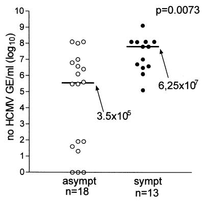
Human cytomegalovirus (HCMV) is the leading cause of congenital viral infection and mental retardation. HCMV infection, while causing asymptomatic infections in most immunocompetent subjects, can be transmitted during pregnancy from the mother with primary (and also recurrent) infection to the fetus. Hence, careful diagnosis of primary infection is required in the pregnant woman based on the most sensitive serologic assays (immunoglobulin M [IgM] and IgG avidity assays) and conventional virologic and molecular procedures for virus detection in blood. Maternal prognostic markers of fetal infection are still under investigation. If primary infection is diagnosed in a timely manner, prenatal diagnosis can be offered, including the search for virus and virus components in fetal blood and amniotic fluid, with fetal prognostic markers of HCMV disease still to be defined. However, the final step for definite diagnosis of congenital HCMV infection is detection of virus in the blood or urine in the first 1 to 2 weeks of life. To date, treatment of congenital infection with antiviral drugs is only palliative both prior to and after birth, whereas the only efficacious preventive measure seems to be the development of a safe and immunogenic vaccine, including recombinant, subunit, DNA, and peptide-based vaccines now under investigation. The following controversial issues are discussed in the light of the most recent advances in the field: the actual perception of the problem; universal serologic screening before pregnancy; the impact of correct counseling on decision making by the couple involved; the role of prenatal diagnosis in ascertaining transmission of virus to the fetus; the impact of preconceptional and periconceptional infections on the prevalence of congenital infection; and the prevalence of congenitally infected babies born to mothers who were immune prior to pregnancy compared to the number born to mothers undergoing primary infection during pregnancy.


Figures

FIG. 1.
FIG. 1.
HCMV replication in human embryonic lung fibroblast cell cultures. (A) HCMV-infected human fibroblast 120 h postinfection (following immunoperoxidase staining with human antibodies), showing intranuclear (IN) and intracytoplasmic (IC) inclusion bodies. (B to D) Electron microscopy of HCMV-infected human fibroblasts. (B) Horseshoe-shaped intranuclear inclusion (IN). (C) Dense bodies (arrows). (D) Maturing virus particles at the level of the nuclear membrane.
FIG. 2.
FIG. 2.
Characteristics of HCMV infection in pregnancy. (From S. Stagno and R. J. Whitley [259], used with permission.)
FIG. 3.
FIG. 3.
Diagram of a longitudinal section that includes a floating and an anchoring chorionic villus at the fetal-maternal interface near the end of the first trimester of human pregnancy. The anchoring villus (AV) functions as a bridge between the fetal and maternal compartments, whereas the floating villus (FV), containing macrophages (M℘, Hofbauer cells) and fetal blood vessels, is bathed by maternal blood. Cytotrophoblasts in the anchoring villus (zone I) form cell columns that attach to the uterine wall (zones II and III). Cytotrophoblasts then invade the uterine interstitium (decidua and first third of the myometrium; zone IV) and maternal vasculature (zone V), thereby anchoring the fetus to the mother and accessing the maternal circulation. Zone designations mark areas in which cytotrophoblasts have distinct patterns of stage-specific antigen expression, including integrin and HLA-G. Decidual granular leukocytes (DGLs) and macrophages (M℘) in maternal blood and fetal capillaries in villous cores are indicated. Areas proposed as sites of natural HCMV transmission to the placenta in utero are numbered 1, 2, and 3. (From Fisher et al. [74], used with permission.)
FIG. 4.
FIG. 4.
(A) Kinetics of IgG, IgM, and neutralizing (Nt) antibody (Ab) response as well as IgG avidity index (AI) in a pregnant woman with primary HCMV infection. (B) Kinetics of infectious virus and different virus products in the blood of the same pregnant woman as in A during the convalescent phase of a primary HCMV infection. Ag, antigenemia; Vir, viremia; DNA, DNAemia; IE mRNA, immediate-early mRNA; +, positive; −, negative; GE, genome equivalents; PBL, peripheral blood leukocytes. (M. G. Revello and G. Gerna, unpublished data.)
FIG. 5.
FIG. 5.
Kinetics of IgM antibody response in 76 pregnant women with primnary HCMV infection as determined in 213 sequential serum samples by using two in-house-developed capture assays in parallel. IgM assays were based on the use of (thin line) virus lysate (213) and (thick line) a commercial recombinant IgM assay (168). (M. G. Revello, G. Gorini, M. Parea, and G. Gerna, unpublished data.)
FIG. 6.
FIG. 6.
Kinetics of IgG avidity index (maturation of HCMV-specific IgG) in 560 serum samples from 176 pregnant women with primary HCMV infection. (M. G. Revello, and G. Gerna, unpublished data.)
FIG. 7.
FIG. 7.
Schematic of diagnosis of primary HCMV infection in pregnancy, including both serologic and virologic approaches. AI, avidity index; Ag, antigenemia; Vir, viremia; DNA, DNAemia; IE mRNA, immediate-early mRNA; NT, neutralization test; Ab, antibody; pos, positive.
FIG. 8.
FIG. 8.
(A) Viremia, indicating the presence in a shell vial monolayer of HCMV p72-positive fibroblast nuclei following cocultivation with peripheral blood leukocytes carrying infectious virus and immunostaining by fluorescein-conjugated p72-specific monoclonal antibody. (B) Antigenemia ex vivo, showing immunofluorescent staining with a pool of monoclonal antibodies of pp65-positive peripheral blood polymorphonuclear leukocytes from a patient with AIDS and disseminated HCMV infection. (C) Antigenemia in vitro, showing pp65-positive polymorphonuclear leukocytes from a healthy blood donor following cocultivation with HCMV-infected human umbilical vein endothelial cells and immunofluorescent staining with the same pool of pp65-specific monoclonal antibodies used in B. (D) Circulating cytomegalic endothelial cell with a pp65-positive leukocyte (arrow). Immunofluorescent staining was done with a pool of pp65-specific monoclonal antibodies.
FIG. 9.
FIG. 9.
Median levels (horizontal lines with values beside arrows) of HCMV antigenemia, viremia, and DNAemia and IgM ratio in fetal blood of symptomatic (sympt) and asymptomatic (asympt) congenitally infected fetuses and newborns. All parameters evaluated were significantly higher in symptomatic than in asymptomatic subjects. Fetuses were considered symptomatic if they had ultrasonographic abnormalities, and newborns were considered symptomatic if they were born with clinical symptoms. PBLs, peripheral blood leukocytes; GE, genome equivalents. (M. G. Revello and G. Gerna, unpublished data.)
FIG. 10.
FIG. 10.
Comparative median levels (horizontal lines with values beside arrows) of DNA in amniotic fluid of mothers with symptomatic (sympt) and asymptomatic (asympt) fetuses. While in both groups all (symptomatic) or most of the (asymptomatic) fetuses showed high levels of HCMV DNA (>105 genome equivalents [GE]/ml), in the asymptomatic group there were seven fetuses, four of whom had DNA levels of <102 genome equivalents/ml and three with undetectable DNA levels at the time of amniocentesis. (M. G. Revello and G. Gerna, unpublished data.)

References

    1. Adler, S. P., S. A. Plotkin, E. Gonczol, M. Cadoz, C. Meric, J. B. Wang, P. Dellamonica, A. M. Best, J. Zahradnik, S. Pincus, K. Berencsi, W. I. Cox, and Z. Gyulai. 1999. A canarypox vector expressing cytomegalovirus (CMV) glycoprotein B primes for antibody responses to a live attenuated CMV vaccine (Towne). J. Infect. Dis. 180:843-846. - PubMed
    1. Ahlfors, K., M. Forsgren, S. A. Ivarsson, S. Harris, and L. Svanberg. 1983. Congenital cytomegalovirus infection: on the relation between type and time of maternal infection and infant's symptoms. Scand. J. Infect. Dis. 15:129-138. - PubMed
    1. Ahlfors, K., S. A. Ivarsson, and H. Nilsson. 1988. On the unpredictable development of congenital cytomegalovirus infection. A study in twins. Early Hum. Dev. 18:125-135. - PubMed
    1. Ahlfors, K., S. A. Ivarsson, S. Harris, L. Svanberg, R. Holmqvist, B. Lernmark, and G. Theander. 1984. Congenital cytomegalovirus infection and disease in Sweden and the relative importance of primary and secondary maternal infections. Preliminary findings from a prospective study. Scand. J. Infect. Dis. 16:129-137. - PubMed
    1. Ahlfors, K., S. A. Ivarsson, and S. Harris. 1999. Report on a long-term study of maternal and congenital cytomegalovirus infection in Sweden. Review of prospective studies available in the literature. Scand. J. Infect. Dis. 31:443-457. - PubMed

Publication types