. 2002 Sep 26;36(1):69-80.
doi: 10.1016/s0896-6273(02)00912-1.

Isomerization and oxidation of vitamin a in cone-dominant retinas: a novel pathway for visual-pigment regeneration in daylight

Affiliations

Isomerization and oxidation of vitamin a in cone-dominant retinas: a novel pathway for visual-pigment regeneration in daylight

Nathan L Mata et al. Neuron. .

Abstract

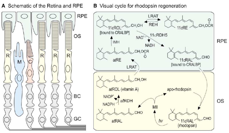
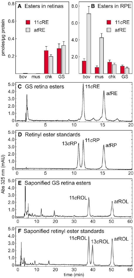
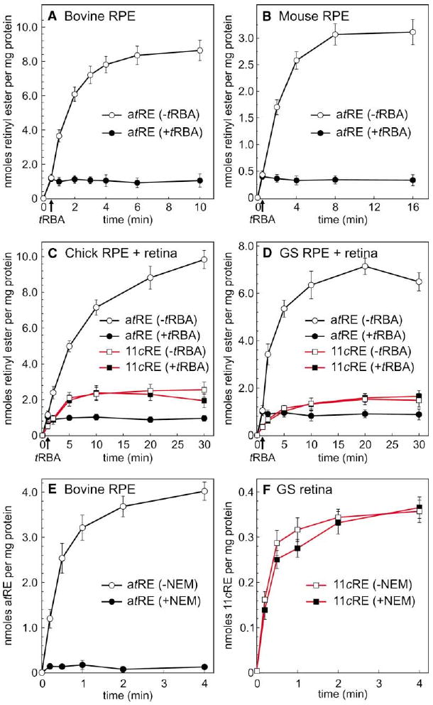
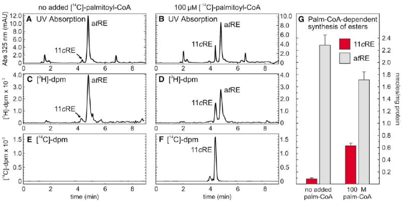
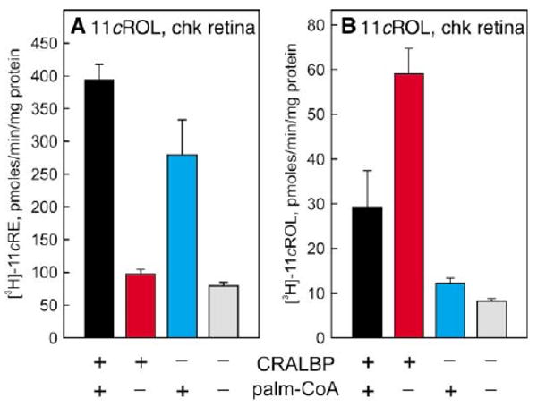
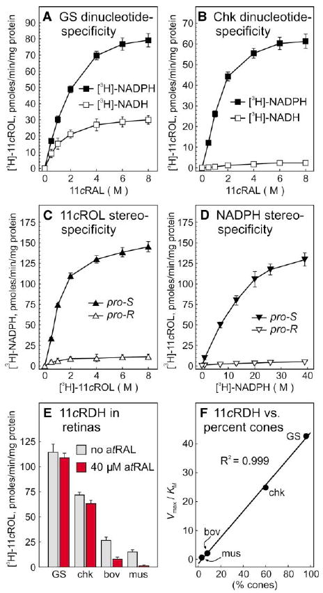
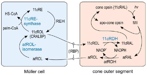
The first step toward light perception is 11-cis to all-trans photoisomerization of the retinaldehyde chromophore in a rod or cone opsin-pigment molecule. Light sensitivity of the opsin pigment is restored through a multistep pathway called the visual cycle, which effects all-trans to 11-cis re-isomerization of the retinoid chromophore. The maximum throughput of the known visual cycle, however, is too slow to explain sustained photosensitivity in bright light. Here, we demonstrate three novel enzymatic activities in cone-dominant ground-squirrel and chicken retinas: an all-trans-retinol isomerase, an 11-cis-retinyl-ester synthase, and an 11-cis-retinol dehydrogenase. Together these activities comprise a novel pathway that regenerates opsin photopigments at a rate 20-fold faster than the known visual cycle. We suggest that this pathway is responsible for sustained daylight vision in vertebrates.


Figures

Figure 1
Figure 1. Organization of the Retina and the Known Visual Cycle
(A) Schematic drawing of a typical vertebrate retina. Rod (R) and cone (C) photoreceptors each contain an outer segment (OS), which is the site of photon capture and the reactions of visual transduction. Adjacent to the outer segments is the retinal pigment epithelium (RPE), which plays a role in the processing of retinoids but is not considered part of the retina. The Müller glial cell (M) may also play a role in retinoid processing. Second-order bipolar cells (BC) and third-order ganglion cells (GC) are shown but not discussed. (B) Visual cycle in rod-dominant retinas. Absorption of a single photon (hv) by a rhodopsin pigment molecule in the rod outer segment induces 11-cis to all-trans isomerization of the 11-cis-retinaldehyde (11cRAL) chromophore to form active metarhodopsin II (MII). MII rapidly decays to yield apo-rhodopsin and free all-trans-retinaldehyde (atRAL). The all-trans-retinaldehyde is reduced to all-trans-retinol (atROL) by all-trans-retinol dehydrogenase (atRDH), which uses NADPH as a cofactor (Lion et al., 1975; Palczewski et al., 1994; Rattner et al., 2000). The all-trans-retinol diffuses across the narrow extracellular space and is taken up by the RPE. Lecithin retinol acyl-transferase (LRAT) catalyzes trans-esterification of a fatty acid from phosphatidylcholine to all-trans-retinol, resulting in formation of an all-trans-retinyl ester (atRE) (Ruiz et al., 1999; Saari and Bredberg, 1989; Shi et al., 1993). Isomerohydrolase (IMH) is postulated to catalyze coupled hydrolysis and isomerization of all-trans-retinyl esters into 11-cis-retinol (11cROL) (Deigner et al., 1989). 11-cis-retinol is oxidized to 11-cis-retinaldehyde by 11-cis-retinol dehydrogenase type-5 (11cRDH5), which uses NAD+ as a cofactor (Lion et al., 1975; Simon et al., 1995). In the dark, under conditions of low 11-cis-retinaldehyde utilization, 11-cis-retinol can also be esterified by LRAT to form 11-cis-retinyl esters (11cRE). Cellular retinaldehyde binding protein (CRALBP) binds 11-cis-retinol and 11-cis-retinaldehyde with high affinity, but not all-trans-retinol (Bunt-Milam and Saari, 1983; Saari and Bredberg, 1987). With chromophore depletion, apo-CRALBP stimulates hydrolysis of 11-cis-retinyl esters by retinyl ester hydrolase (REH) (Stecher et al., 1999). 11-cis-retinaldehyde diffuses across the extracellular space, is taken up by the outer segment, and recombines with apo-rhodopsin to regenerate rhodopsin.
Figure 2
Figure 2. Retinyl Esters in Retina and RPE
(A) Levels of 11-cis-retinyl esters (11cRE, red bars) and all-trans-retinyl esters (atRE, gray bars) in bovine (bov), mouse (mus), chicken (chk), and ground squirrel (GS) retinas. (B) Levels of 11-cis-retinyl esters and all-trans-retinyl esters in RPE from the four species. Values are shown as pmols per μg protein in microsomal membranes. Error bars show standard deviations (n = 4). (C) Representative HPLC chromatogram of retinyl esters from ground squirrel retina. (D) Representative HPLC chromatogram of the retinyl-ester standards: 13-cis-retinyl palmitate (13cRP), 11-cis-retinyl palmitate (11cRP), and all-trans-retinyl palmitate (atRP). (E) Representative chromatogram of saponified retinyl esters from GS retina showing 11-cis-retinol (11cROL) and all-trans-retinol (atROL). (F) Representative chromatogram of saponified retinyl-ester standards showing 11-cis-retinol, 13-cis-retinol (13cROL), and all-trans-retinol. UV absorption at 325 nm is shown in milliabsorption units (mAU).
Figure 3
Figure 3. Retinyl Esters Synthesized by RPE and Retinal Membranes from All-trans-Retinol
(A) Time course of all-trans-retinyl ester (atRE) synthesis by bovine RPE membranes in the absence (open circles) or presence (filled circles) of tRBA inhibitor. Arrow on the baseline indicates the time of tRBA addition. (B) Time course of all-trans-retinyl ester synthesis by mouse RPE membranes in the absence or presence of tRBA inhibitor. (C) Time course of all-trans-retinyl ester (circles, black lines) or 11-cis-retinyl ester (11cRE, squares, red lines) synthesis by chicken combined RPE + retinal membranes in the absence (open figures) or presence (filled figures) of tRBA inhibitor. (D) Time course of all-trans-retinyl ester or 11-cis-retinyl ester synthesis by ground-squirrel combined RPE + retinal membranes in the absence or presence of tRBA inhibitor. (E) Time course of all-trans-retinyl ester synthesis by bovine RPE membranes plus (open circles) or minus (filled circles) pre-treatment with NEM. (F) Time course of 11-cis-retinyl ester synthesis by ground-squirrel retinal membranes plus (open squares) or minus (filled squares) pre-treatment with NEM. For each experiment, the indicated all-trans- and 11-cis-retinyl esters were the only retinyl esters formed, shown as nmols per mg protein. Error bars show standard deviations (n = 4).
Figure 4
Figure 4. Synthesis of 11-cis-Retinyl Esters by Ground-Squirrel RPE + Retinal Membranes Is Stimulated by Palm-CoA
[3H]-all-trans-retinol, plus or minus [14C]-palm-CoA, were added to UV-treated, combined RPE + retinal membranes from ground squirrel. Following incubation for 40 min, extracted retinoids were resolved by HPLC with simultaneous UV and online-radiometric analysis. Chromatograms are shown for 325 nm absorption (A and B), [3H]-decays per minute (dpm) (C and D), and [14C]-dpm (E and F). Note that [14C]-palmitate is incorporated into 11-cis-retinyl esters (11cRE) but not all-trans-retinyl esters (atRE). (G) Histogram showing levels of 11-cis-retinyl esters (red bars) and all-trans-retinyl esters (gray bars) synthesized during a 5 min incubation in the absence or presence of palm-CoA, in nmols per mg protein. Error bars show standard deviations (n = 4).
Figure 5
Figure 5. Effects of apo-CRALBP on the Synthesis of 11-cis-Retinyl Esters and 11-cis-Retinol by Chicken Retinal Membranes
(A) Synthesis of [3H]-11-cis-retinyl esters (11cRE) from [3H]-all-trans-retinol by chicken retinal membranes in the presence of 30 μM apo-CRALBP (black and red bars), 100 μM palm-CoA (black and cyan bars), or absence of both (gray bar). Shown as specific activities (pmols [3H]-11-cis-retinyl esters per min per mg protein). (B) Synthesis of [3H]-11-cis-retinol (11cROL) from [3H]-all-trans-retinol by ground-squirrel retinal membranes in the presence of apo-CRALBP, palm-CoA, or absence of both. Shown as specific activities (pmols [3H]-11-cis-retinol per min per mg protein). Error bars show standard deviations (n = 4). Note the palm-CoA dependence of 11-cis-retinyl ester synthesis and the apo-CRALBP dependence of 11-cis-retinol synthesis.
Figure 6
Figure 6. 11-cis-Retinol Dehydrogenase Activity in Ground-Squirrel and Chicken Retinas
Microsomal membranes from ground squirrel (A) and chicken (B) retinas were assayed for the capacity to reduce 11-cis-retinaldehyde (11cRAL) to [3H]-11-cis-retinol (11cROL) in the presence of [3H]-NADH (open squares) or [3H]-NADPH (filled squares). 11-cis-retinol dehydrogenase (11cRDH) activity is expressed in pmols [3H]-11-cis-retinol per min per mg protein. (C) Ground-squirrel retinal microsomes were assayed for the capacity to synthesize [3H]-NADPH from pro-R- or pro-S-[3H]-11-cis-retinol plus unlabeled NADP+. (D) Ground-squirrel retinal microsomes were assayed for the capacity to synthesize [3H]-11-cis-retinol from pro-R- or pro-S-[3H]-NADPH. (E) Microsomes from ground squirrel (GS), chicken (chk), bovine (bov), and mouse (mus) retinas were assayed for the capacity to synthesize [3H]-11-cis-retinol from 11-cis-retinaldehyde and [3H]-NADPH in the absence (gray bars) or presence (red bars) of 40 μM all-trans-retinaldehyde (atRAL). Error bars show standard deviations (n = 4). (F) Derived Vmax/KM (catalytic efficiencies) for 11-cis-retinol dehydrogenase in mouse, bovine, chicken, and ground-squirrel retinas are plotted against the percentage of cone photoreceptors in each species. Linear regression analysis revealed a correlation coefficient (R2) of 0.999.
Figure 7
Figure 7. Proposed Visual Cycle for Regeneration of Cone Opsin
Absorption of a photon (hv) induces 11-cis to all-trans isomerization of the retinaldehyde chromophore, resulting in activated opsin (MII) inside a cone outer segment. Decay of MII releases all-trans-retinaldehyde (atRAL), which is reduced to all-trans-retinol (atROL) by NADPH-dependent all-trans-retinol dehydrogenase (atRDH). all-trans-retinol released into the extracellular space by rods and cones is taken up by Müller cells, which contain the novel all-trans-retinol isomerase activity. This enzyme catalyzes passive isomerization between all-trans-retinol and 11-cis-retinol. Müller cells also contain CRALBP, which binds 11-cis-retinol but not all-trans-retinol (Bunt-Milam and Saari, 1983). Isomerization of all-trans-retinol to 11-cis-retinol (11cROL) is driven by mass-action through the activity of a novel 11-cis-retinyl ester (11cRE) synthase, which catalyzes esterification of 11-cis-retinol using palm-CoA (or other fatty-acyl-CoA's) as an acyl-donor. Retinyl ester hydrolase (REH) is activated by apo-CRALBP to yield 11-cis-retinol, which is released by the Müller cell and taken up by cones. The all-trans-retinol and 11-cis-retinol within the extracellular space in transit between cones and Müller cells are bound to IRBP. Within the cone outer segment is a new NADP+-dependent 11-cis-retinol dehydrogenase (11cRDH), which oxidizes 11-cis-retinol to 11-cis-retinaldehyde. 11-cis-retinaldehyde combines with apo-opsin to regenerate cone opsin pigment. The novel enzymatic activities presented in this paper are displayed in blue.

Comment in

References

    1. Adler AJ, Spencer SA. Effect of light on endogenous ligands carried by interphotoreceptor retinoid-binding protein. Exp Eye Res. 1991;53:337–346. - PubMed
    1. Baylor DA, Lamb TD, Yau KW. The membrane current of single rod outer segments. J Physiol. 1979;288:589–611. - PMC - PubMed
    1. Baylor DA, Nunn BJ, Schnapf JL. The photocurrent, noise and spectral sensitivity of rods of the monkey Macaca fascicularis. J Physiol. 1984;357:575–607. - PMC - PubMed
    1. Bok D, Ong DE, Chytil F. Immunocytochemical localization of cellular retinol binding protein in the rat retina. Invest Ophthalmol Vis Sci. 1984;25:877–883. - PubMed
    1. Bunt-Milam AH, Saari JC. Immunocytochemical localization of two retinoid-binding proteins in vertebrate retina. J Cell Biol. 1983;97:703–712. - PMC - PubMed

Publication types

MeSH terms