Immunohistochemical localization of feline immunodeficiency virus using native species antibodies
- PMID: 12368188
- PMCID: PMC1867283
- DOI: 10.1016/S0002-9440(10)64391-X
Immunohistochemical localization of feline immunodeficiency virus using native species antibodies
Abstract
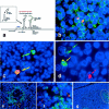
Feline immunodeficiency virus (FIV) is the feline analog of human immunodeficiency virus and a small animal model of human acquired immune deficiency syndrome (AIDS). We sought to identify early in vivo target cells in cats infected with clade B or C FIV. In tissues, however, neither mouse monoclonal nor rabbit polyclonal antibodies suitably detected FIV because of either insensitivity or lack of specificity. We therefore developed an immunohistochemical protocol using high-antibody-titer serum from cats chronically infected with FIV(Petaluma). Native species anti-FIV antibodies were labeled with biotinylated protein A before placement on tissues, and downstream signal was tyramide-amplified. This method revealed many productively infected cells in bone marrow, lymph node, thymus, mucosal-associated lymphoid tissue, and spleen, but few such cells in liver and none in kidney or brain. Concurrent labeling for virus and cell phenotype revealed that antigen-bearing populations were primarily T lymphocytes but included macrophages and dendritic cells. Our results demonstrate that FIV: 1) expands rapidly in T cells, 2) targets long-lived reservoir populations, and 3) is replicatively quiescent in brain at 3 weeks after infection. Use of native species antibodies for immunohistochemical detection of infectious antigens has application to other settings in which xenotypic (eg, mouse and rabbit) antibody sources are inadequate or unavailable.
Figures


Similar articles
-
Selective thymocyte depletion and immunoglobulin coating in the thymus of cats infected with feline immunodeficiency virus.AIDS Res Hum Retroviruses. 1997 May 1;13(7):611-20. doi: 10.1089/aid.1997.13.611. AIDS Res Hum Retroviruses. 1997. PMID: 9135879
-
Localization of the viral antigen of feline immunodeficiency virus in the lymph nodes of cats at the early stage of infection.Arch Virol. 1993;131(3-4):335-47. doi: 10.1007/BF01378636. Arch Virol. 1993. PMID: 8102229
-
In vivo infection of ramified microglia from adult cat central nervous system by feline immunodeficiency virus.Virology. 2000 Mar 15;268(2):420-9. doi: 10.1006/viro.1999.0152. Virology. 2000. PMID: 10704350
-
Feline immunodeficiency virus: an interesting model for AIDS studies and an important cat pathogen.Clin Microbiol Rev. 1995 Jan;8(1):87-112. doi: 10.1128/CMR.8.1.87. Clin Microbiol Rev. 1995. PMID: 7704896 Free PMC article. Review.
-
Central and peripheral reservoirs of feline immunodeficiency virus in cats: a review.J Gen Virol. 2017 Aug;98(8):1985-1996. doi: 10.1099/jgv.0.000866. Epub 2017 Aug 28. J Gen Virol. 2017. PMID: 28749325 Review.
Cited by
-
Peripheral and central immune cell reservoirs in tissues from asymptomatic cats chronically infected with feline immunodeficiency virus.PLoS One. 2017 Apr 6;12(4):e0175327. doi: 10.1371/journal.pone.0175327. eCollection 2017. PLoS One. 2017. PMID: 28384338 Free PMC article.
-
FIV establishes a latent infection in feline peripheral blood CD4+ T lymphocytes in vivo during the asymptomatic phase of infection.Retrovirology. 2012 Feb 7;9:12. doi: 10.1186/1742-4690-9-12. Retrovirology. 2012. PMID: 22314004 Free PMC article.
-
Intracellular nucleotide levels and the control of retroviral infections.Virology. 2013 Feb 20;436(2):247-54. doi: 10.1016/j.virol.2012.11.010. Epub 2012 Dec 20. Virology. 2013. PMID: 23260109 Free PMC article. Review.
-
Prior virus exposure alters the long-term landscape of viral replication during feline lentiviral infection.Viruses. 2011 Oct;3(10):1891-908. doi: 10.3390/v3101891. Epub 2011 Oct 13. Viruses. 2011. PMID: 22069521 Free PMC article.
-
Viral Reservoirs in Lymph Nodes of FIV-Infected Progressor and Long-Term Non-Progressor Cats during the Asymptomatic Phase.PLoS One. 2016 Jan 7;11(1):e0146285. doi: 10.1371/journal.pone.0146285. eCollection 2016. PLoS One. 2016. PMID: 26741651 Free PMC article.
References
-
- Haase AT, Henry K, Zupancic M, Sedgewick G, Faust RA, Melroe H, Cavert W, Gebhard K, Staskus K, Zhang ZQ, Dailey PJ, Balfour HH, Jr, Erice A, Perelson AS: Quantitative image analysis of HIV-1 infection in lymphoid tissue. Science 1996, 274:985-989 - PubMed
-
- Zhang ZQ, Notermans DW, Sedgewick G, Cavert W, Wietgrefe S, Zupancic M, Gebhard K, Henry K, Boies L, Chen Z, Jenkins M, Mills R, McDade H, Goodwin C, Schuwirth CM, Danner SA, Haase AT: Kinetics of CD4+ T cell repopulation of lymphoid tissues after treatment of HIV-1 infection. Proc Natl Acad Sci USA 1998, 95:1154-1159 - PMC - PubMed
-
- Embretson J, Zupancic M, Ribas JL, Burke A, Racz P, Tenner-Racz K, Haase AT: Massive covert infection of helper T lymphocytes and macrophages by HIV during the incubation period of AIDS. Nature 1993, 362:359-362 - PubMed
-
- Schacker T, Little S, Connick E, Gebhard-Mitchell K, Zhang ZQ, Krieger J, Pryor J, Havlir D, Wong JK, Richman D, Corey L, Haase AT: Rapid accumulation of human immunodeficiency virus (HIV) in lymphatic tissue reservoirs during acute and early HIV infection: implications for timing of antiretroviral therapy. J Infect Dis 2000, 181:354-357 - PubMed
Publication types
MeSH terms
Substances
Grants and funding
LinkOut - more resources
Full Text Sources
Other Literature Sources
Miscellaneous

