

Genetic changes in neoplasms arising in congenital melanocytic nevi: differences between nodular proliferations and melanomas
- PMID: 12368190
- PMCID: PMC1867277
- DOI: 10.1016/S0002-9440(10)64393-3
Genetic changes in neoplasms arising in congenital melanocytic nevi: differences between nodular proliferations and melanomas
Abstract
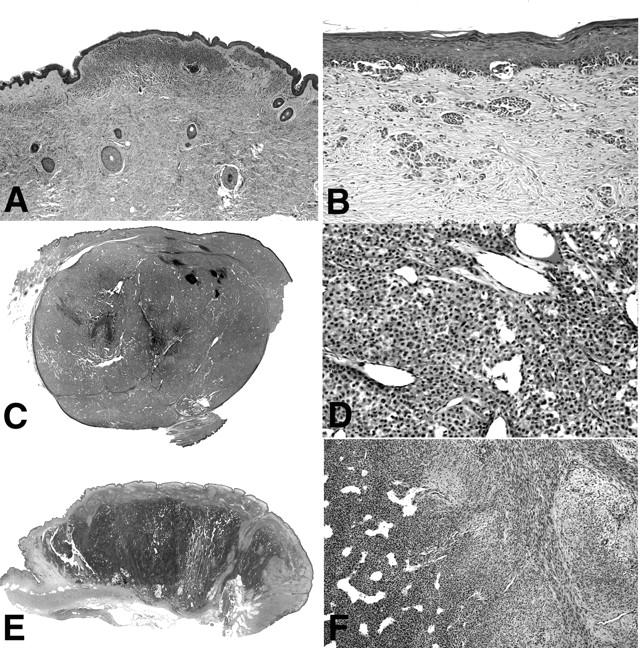
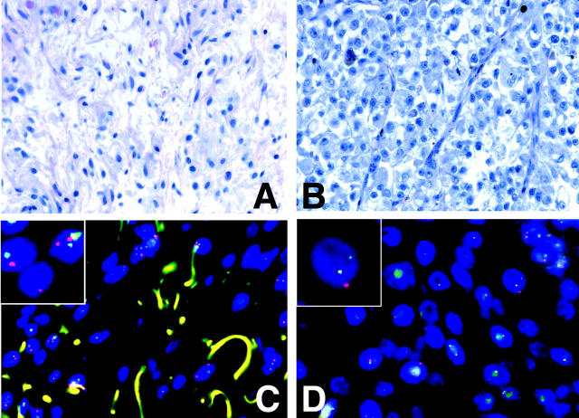
Large congenital melanocytic nevi (CMN) are at an increased risk of developing melanoma. Several forms of secondary proliferations can arise in congenital nevi on rare occasions. Although some of these closely resemble melanoma both clinically and histologically, metastasis is rare. We used comparative genomic hybridization to analyze chromosomal aberrations in different types of proliferations arising in CMN and compared them to typical congenital nevi, clear-cut melanomas arising in congenital nevi, as well as primary cutaneous melanomas that were not associated with a CMN. Cases of CMN and CMN with secondary proliferations were assigned to six groups according to the predominant histological pattern: group I, bland congenital nevi (n = 6); group II, congenital nevi with foci of increased cellularity (n = 4); group III, CMN with a proliferation simulating superficial spreading melanoma in situ (n = 3); group IV, CMN with a proliferation simulating nodular melanoma (n = 9); group V, proliferating neurocristic hamartoma (n = 1); and group VI, melanoma arising in congenital nevus (n = 6). No aberrations were found in groups I to III, whereas seven of nine cases of group IV, and one of one case of group V, showed aberrations. In group IV six of seven cases with aberrations (86%) showed numerical aberrations of whole chromosomes exclusively. This pattern differed significantly from the findings in melanoma that arose within CMN (n = 6), group VI, or independent of CMN (n = 122) in which only 5% showed numerical changes only. The single case in group V showed aberrations similar to melanoma. The finding of frequent numerical chromosomal aberrations in atypical nodular proliferations arising in CMN identifies these as clonal neoplasms with a genomic instability consistent with a mitotic spindle checkpoint defect. This difference compared to the aberration pattern found in melanoma might explain their more benign clinical behavior and may be of diagnostic value in ambiguous cases.
Figures
References
-
- Rhodes AR: Melanocytic precursors of cutaneous melanoma. Estimated risks and guidelines for management. Med Clin North Am 1986, 70:3-37 - PubMed
-
- Sahin S, Levin L, Kopf AW, Rao BK, Triola M, Koenig K, Huang C, Bart R: Risk of melanoma in medium-sized congenital melanocytic nevi: a follow-up study. J Am Acad Dermatol 1998, 39:428-433 - PubMed
-
- Swerdlow AJ, English JS, Qiao Z: The risk of melanoma in patients with congenital nevi: a cohort study. J Am Acad Dermatol 1995, 32:595-599 - PubMed
-
- Ruiz-Maldonado R, Tamayo L, Laterza AM, Duran C: Giant pigmented nevi: clinical, histopathologic, and therapeutic considerations. J Pediatr 1992, 120:906-911 - PubMed
-
- Quaba AA, Wallace AF: The incidence of malignant melanoma (0 to 15 years of age) arising in “large” congenital nevocellular nevi. Plast Reconstr Surg 1986, 78:174-181 - PubMed
Publication types
MeSH terms
Substances
LinkOut - more resources
Full Text Sources
Medical
