. 2002 Oct;161(4):1187-98.
doi: 10.1016/S0002-9440(10)64395-7.

Heterogeneity of ductular reactions in adult rat and human liver revealed by novel expression of deleted in malignant brain tumor 1

Affiliations

Heterogeneity of ductular reactions in adult rat and human liver revealed by novel expression of deleted in malignant brain tumor 1

Hanne Cathrine Bisgaard et al. Am J Pathol. 2002 Oct.

Abstract

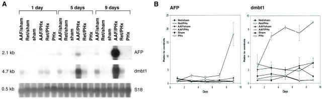
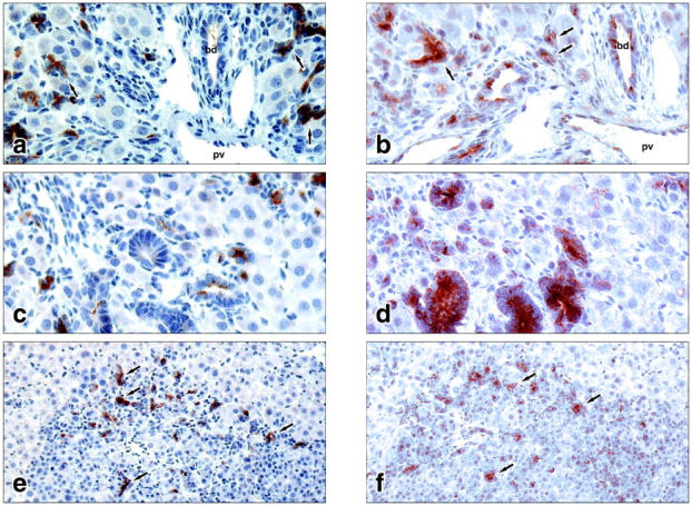
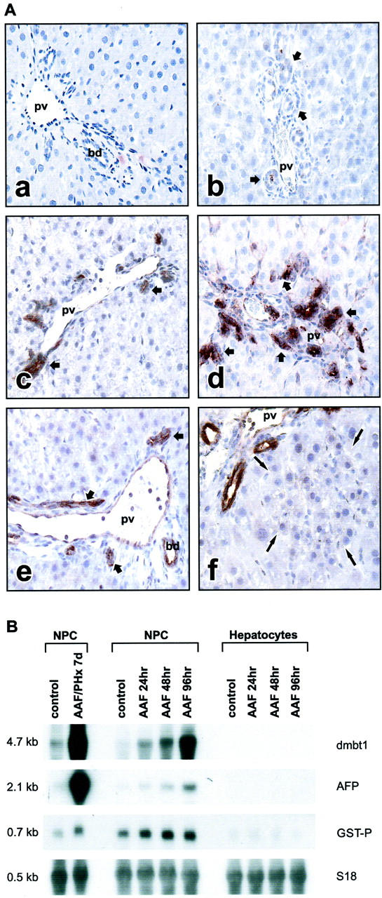
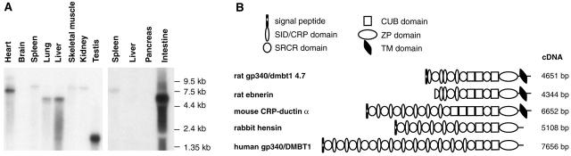
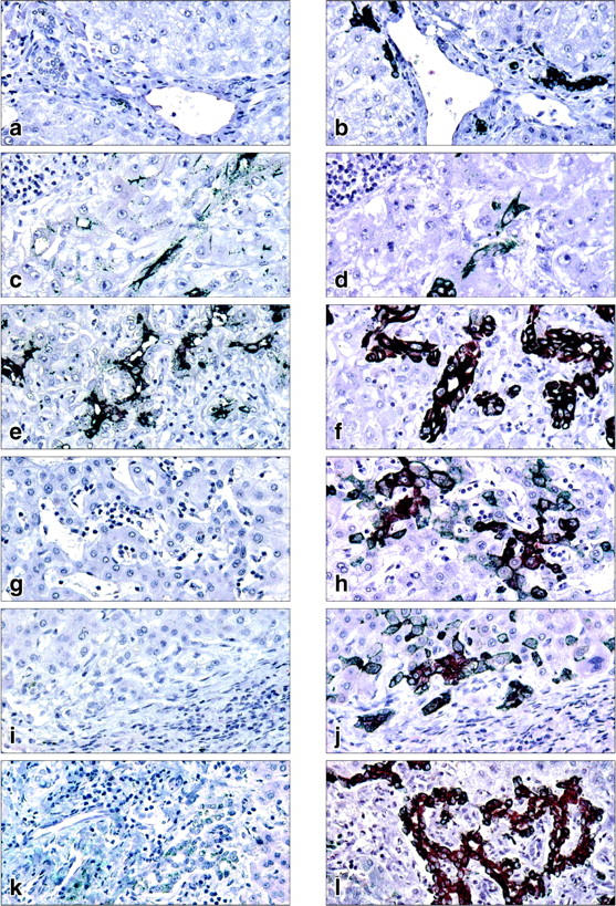
The regenerative capacity of mammalian adult liver reflects the ability of a number of cell populations within the hepatic lineage to take action. Limited information is available regarding factors and mechanisms that determine the specific lineage level at which liver cells contribute to liver repair as well as the fate of their progeny in the hostile environment created by liver injury. In the present study, we attempted to identify novel molecules preferentially involved in liver regeneration by recruitment of transit-amplifying, ductular (oval) cell populations. With a subtractive cDNA library screening approach, we identified 48 enriched, nonredundant gene products associated with liver injury and oval cell proliferation in the adult rat liver. Of these, only two, namely alpha-fetoprotein and a novel transcript with high homology to human DMBT1 (deleted in malignant brain tumor 1), were specifically associated with the emergence of ductular (oval) cell populations in injured liver. Subsequent cloning and characterization of the rat DMBT1 homologue revealed a highly inducible expression in ductular reactions composed of transit-amplifying ductular (oval) cells, but not in ductular reactions after ligation of the common bile duct. In human liver diseases, DMBT1 was expressed in ductular reactions after infection with hepatitis B and acetaminophen intoxication, but not in primary biliary cirrhosis, primary sclerosing cholangitis, and obstruction of the large bile duct. The expression heterogeneity in ductular reactions and multiple functions of DMBT1 homologues point to intriguing roles in regulating not only tissue repair but also fate decision and differentiation paths of specific cell populations in the hepatic lineage.


Figures

Figure 1.
Figure 1.
Gene expression analysis of AFP and the DMBTt1 homologue in rat liver. A: Northern blot analysis. Each lane contains 10 μg of total RNA isolated from one rat liver. Membranes were hybridized to cDNA probes labeled with [32P]dCTP and visualized by autoradiography. Hybridization with S18 was used to assess integrity and equal loading of RNA samples. B: Quantification of gene expression by slot blot and phosporimager analysis. Each point represents the mean of hepatic gene expression in three animals normalized to the expression in a control group of three untreated rats. AAF/sham, treatment with 2-AAF and laparotomy only; Ret/sham, treatment with retrorsine and laparotomy only; sham, laparotomy only; AAF/PHx, treatment with 2-AAF and a 70% hepatectomy; Ret/PHx, treatment with retrorsine and a 70% hepatectomy; PHx, 70% hepatectomy only. Animals were sacrificed 1, 3, 5, 7, and 9 days after the surgical procedure.
Figure 2.
Figure 2.
Cellular localization of AFP (a, c, e) and the rat homologue of DMBT1 (b, d, f) at day 9 after a 70% hepatectomy in the AAF/PHx protocol. a and b feature a portal area with an intricate network of transit-amplifying ductular (oval) cells penetrating the hepatic parenchyma. c and d feature structures of intestinal-type tissue staining positive for dmbt1 but not for AFP. e and f feature a focus of small basophilic hepatocytes entrapping structures of ductular (oval) cells staining positive for both AFP and dmbt1. pv, portal vein; bd, bile duct; arrows point to structures of ductular (oval) cells. Original magnifications: ×400 (c and d); ×200 (e and f).
Figure 3.
Figure 3.
A: Localization of rat dmbt1 in normal rat liver (a), 5 days after ligation of the common bile duct (b), 5 days after initiation of 2-AAF treatment (c), 5 days after partial hepatectomy in the AAF/PHx protocol (d), 5 days after partial hepatectomy (e), and 5 days after partial hepatectomy in the Ret/PHx model (f). Short arrows point to ductular structures; long arrows point to a focus of small hepatocyte-like cells. pv, portal vein; bd, bile duct. B: Analysis of dmbt1 and AFP transcripts in isolated liver cell populations by Northern blotting of 10 μg of total RNA. Hybridization to the placental form of glutathione S-transferase (GST-P) was used as a common marker for bile duct and ductular (oval) cells to verify the purity of nonparenchymal (NPC) versus parenchymal (hepatocytes) cell populations. Hybridization with S18 was used to assess integrity and equal loading of RNA. Original magnifications, ×200 (a to f).
Figure 4.
Figure 4.
A: Northern blot analysis of DMBT1 homologues in normal adult rat tissues. Left: Samples of 2 μg of poly (A+) RNA are loaded in each lane and the membrane exposed to X-ray film for 7 days. Right: Samples of 10 μg of total RNA are loaded in each lane and the membrane exposed to X-ray film for 2 days. B: Domain organization of rat dmbt1 4.7kb and members of the SRCR superfamily group B with high homology to dmbt1 4.7kb.
Figure 5.
Figure 5.
Representative immunostaining of DMBT1 (a, c, e, g, i, k) or cytokeratin 7 (CK7) (b, d, f, h, j, l) in human liver disease with ductular reactions. Pairs of serial sections were used in the analysis. Normal liver (a, b), hepatitis B infection (c, d), acetaminophen intoxication (e, f), primary biliary cirrhosis (g, h), primary sclerosing cholangitis (i, j), and large bile duct obstruction (k, l). Original magnifications, ×400.

Comment in

Similar articles

Cited by

References

    1. Blau HM, Brazelton TR, Weimann JM: The evolving concept of a stem cell: entity or function. Cell 2001, 105:829-841 - PubMed
    1. Michalopoulos GK, De Frances MC: Liver regeneration. Science 1997, 276:60-66 - PubMed
    1. Overturf K, Al-Dhalimy M, Ou C, Grompe M: Serial transplantation reveals the stem-cell-like regenerative potential of adult mouse hepatocytes. Am J Pathol 1997, 51:1273-1280 - PMC - PubMed
    1. Lemire JM, Shiojiri N, Fausto N: Oval cell proliferation and the origin of small hepatocytes in liver injury induced by D-galactosamine. Am J Pathol 1991, 139:535-552 - PMC - PubMed
    1. Dabeva MD, Shafritz D: Activation, proliferation, and differentiation of progenitor cells into hepatocytes in the D-galactosamine model of liver regeneration. Am J Pathol 1993, 43:1606-1620 - PMC - PubMed

Publication types

MeSH terms