. 2002 Oct;161(4):1255-63.
doi: 10.1016/s0002-9440(10)64402-1.

Virus-induced demyelination in nude mice is mediated by gamma delta T cells

Affiliations

Virus-induced demyelination in nude mice is mediated by gamma delta T cells

Ajai A Dandekar et al. Am J Pathol. 2002 Oct.

Abstract

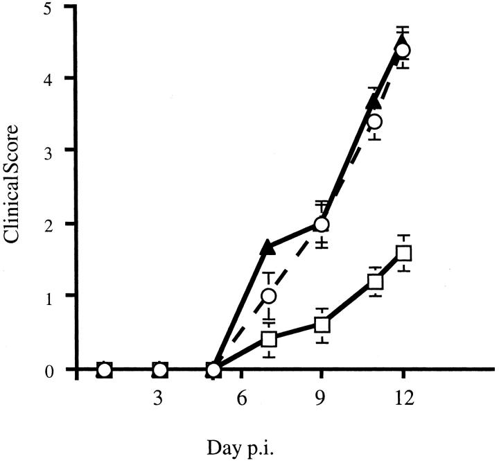
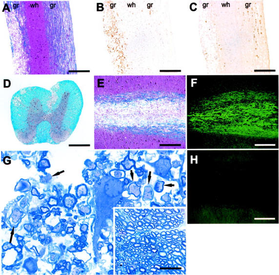
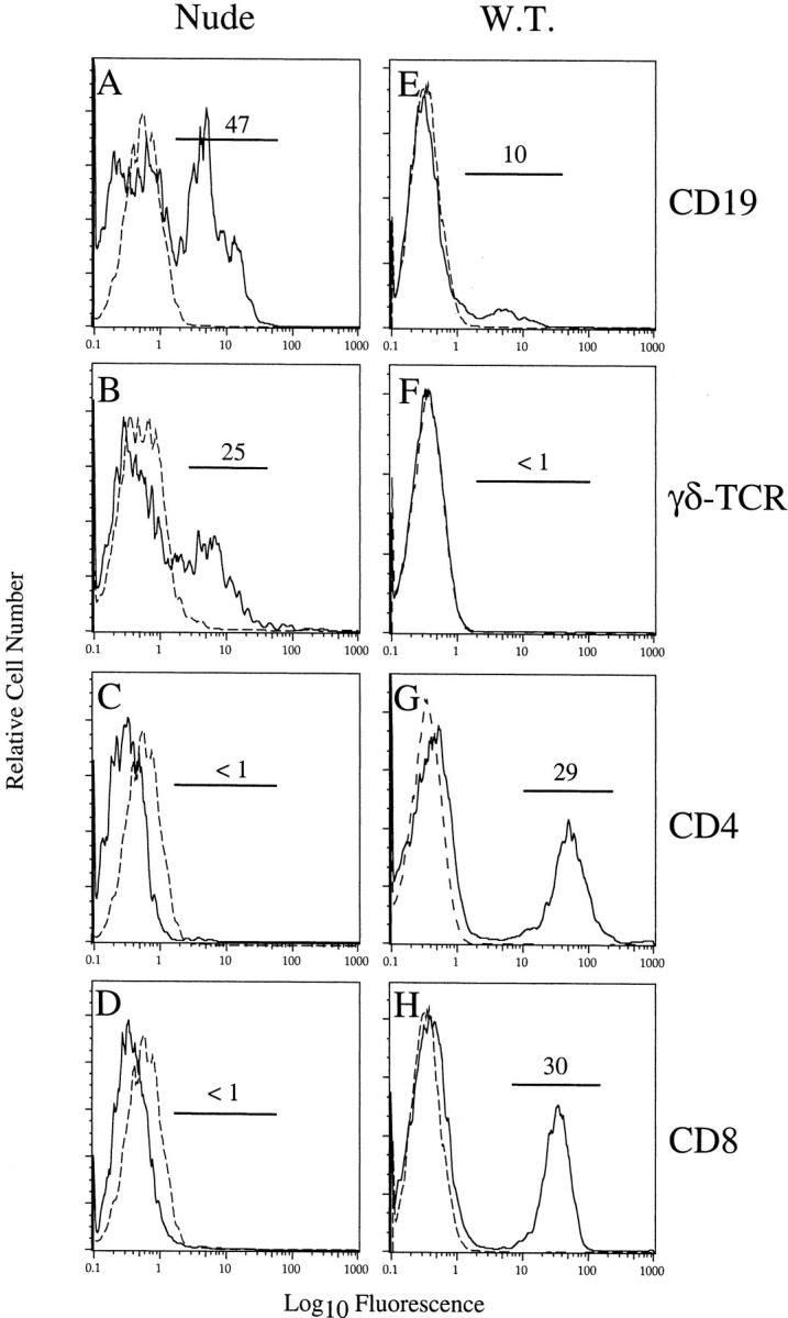
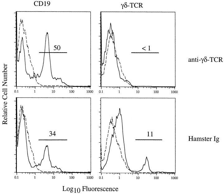
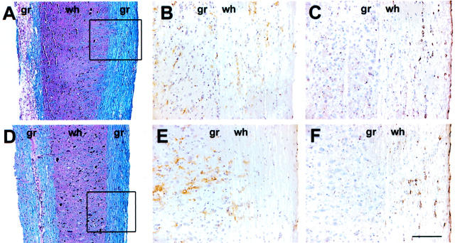
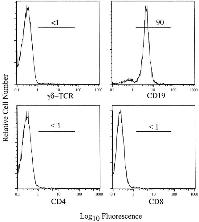
Infection of mice with mouse hepatitis virus (MHV), strain JHM, results in acute and chronic demyelination with many similarities to the human disease multiple sclerosis. This pathological process is primarily T cell-mediated and MHV infection of mice lacking B and T cells does not result in demyelination. In apparent contradiction to these results, robust demyelination is detected in MHV-infected young nude (athymic) mice. Herein, we show that demyelination in nude mice was mediated by gamma delta T cells. These cells, but not conventional CD4 or CD8 alpha beta T cells, were detected in the central nervous system of MHV-infected nude mice and their depletion with neutralizing antibody resulted in an 80% reduction in demyelination. These results show, for the first time, that gamma delta T cells can substitute for alpha beta T cells in a virus model of demyelination and further support a pathological role for gamma delta T cells in patients with multiple sclerosis.


Figures

Figure 1.
Figure 1.
Clinical disease in MHV-infected nude mice. BALB/c-nude mice infected with MHV were evaluated for clinical disease. Untreated mice (filled triangles) and those treated with hamster Ig (open circles) demonstrated similar disease progression. All untreated mice died by day 13 after inoculation. Mice treated with anti-γδ mAb UC7-13D5 (open squares) exhibited significantly less severe disease. Five mice per group were analyzed. Disease scoring scale: 0, asymptomatic; 1, limp tail; 2, wobbly gait; 3, hindlimb paresis; 4, quadriparesis/paralysis; 5, moribund/dead.
Figure 2.
Figure 2.
Detection of demyelination in MHV-infected BALB/c-nude mice at 12 days after inoculation. Spinal cords were harvested and 8-μm sagittal sections were stained for demyelination (A), macrophages (B), or viral antigen (C) as described in Materials and Methods. Demyelination was detected (A) in an area of macrophage/microglia infiltration (B). C: Viral antigen was abundant in normal appearing white matter and diminished in areas of frank demyelination. D: Demyelination was localized primarily in the ventral and ventrolateral funiculi with lesser amounts in the dorsal white matter tracts. In areas of gross demyelination (E), axons are preserved (F) as indicated by staining with anti-phosphoneurofilament H Ab. H: No staining was detected in the absence of primary antibody. G: Toluidine blue staining of epon-embedded sections revealed many axons with diminished or absent myelin staining (arrows). The inset in G shows an area of normal myelin from the spinal cord of the same mouse. g, Gray matter; wh, white matter. Scale bars: 100 μm (A–D); 50 μm (E, F, H); 20 μm (G); 40 μm (inset in G).
Figure 3.
Figure 3.
B and γδ T cells, but not CD4 or CD8 T cells, were detected in the CNS of MHV-infected BALB/c-nude mice. Lymphocytes were harvested from the CNS of BALB/c-nude (A–D) or BALB/c (E–H) mice at 12 days after inoculation. Lymphocytes from three BALB/c-nude mice were pooled and stained for flow cytometry as described in Materials and Methods. Cells from individual BALB/c mice were similarly analyzed. Although B cells were present in both samples (A, E), γδ T cells were detected only in the CNS of MHV-infected nude mice (B, F). No CD4 or CD8 T cells, including CD8αα T cells, were present in the CNS of MHV-infected nude mice (C, D). CD4 and CD8 T cells comprised 60% of mononuclear cells identified in the CNS of MHV-infected BALB/c mice (G, H). Dashed lines represent staining with fluorophore-conjugated control antibody. Results shown are representative of three independent experiments.
Figure 4.
Figure 4.
Depletion of γδ T cells from infected BALB/c-nude mice. Nude mice were treated with anti-γδ-TCR mAb UC7-13D5 or hamster Ig as a control as described in Materials and Methods. These mice were then infected intracranially with MHV and 12 days later lymphocytes were harvested from the CNS and examined by flow cytometry. No γδ T cells were detected in the CNS after treatment with anti-γδ-TCR mAb but were readily detectable after hamster Ig treatment. Treatment with either antibody did not substantially affect the frequency of B cells in the infected CNS. Dashed lines represent staining with fluorophore-conjugated Ig. The results shown are representative of three independent experiments.
Figure 5.
Figure 5.
Depletion of γδ T cells altered the number and location of macrophages/microglia in the spinal cord. Serial sections from spinal cords of hamster Ig-treated (A–C) or anti-γδ-TCR mAb-treated (D–F) BALB/c-nude mice were stained for myelin (A, D), macrophages/microglia (B, E), or viral antigen (C, F). Extensive demyelination was observed only in hamster Ig-treated sections (A, D). Distribution of macrophages/microglia and virus antigen was examined in an area of intact white matter with minimal demyelination (boxes in A and D). Macrophages/microglia infiltrated the white matter of hamster Ig-treated mice (B), whereas in anti-γδ-TCR mAb-treated mice these cells were predominately in the gray matter (E), despite the presence of equivalent amounts of virus in both spinal cords (C, F). gr, Gray matter; wh, white matter. Scale bars: 100 μm (A, D); 50 μm (B, C, E, F).
Figure 6.
Figure 6.
B cells comprised the great majority of cells in the spleen of nude mice 7 days after intraperitoneal inoculation with MHV. Splenocytes were prepared from MHV-infected B6-nude mice and analyzed by flow cytometry after staining for CD4, CD8, CD19, and γδ TCR. The vast majority of cells were CD19+ B cells with few γδ, CD4, or CD8 T cells detected.

Similar articles

Cited by

References

    1. Hemmer B, Archelos JJ, Hartung HP: New concepts in the immunopathogenesis of multiple sclerosis. Nat Rev Neurosci 2002, 3:291-301 - PubMed
    1. Noseworthy JH, Lucchinetti C, Rodriguez M, Weinshenker BG: Multiple sclerosis. N Engl J Med 2000, 343:938-952 - PubMed
    1. Murray PD, Pavelko KD, Leibowitz J, Lin X, Rodriguez M: CD4(+) and CD8(+) T cells make discrete contributions to demyelination and neurologic disease in a viral model of multiple sclerosis. J Virol 1998, 72:7320-7329 - PMC - PubMed
    1. Tsunoda I, Fujinami RS: Two models of multiple sclerosis: experimental allergic encephalomyelitis and Theiler’s murine encephalomyelitis virus. J Neuropathol Exp Neurol 1996, 55:672-686 - PubMed
    1. Steinman L: Assessment of animal models for MS and demyelinating disease in the design of rational therapy. Neuron 1999, 24:511-514 - PubMed

Publication types

MeSH terms

Substances