. 2002 Oct;161(4):1419-27.
doi: 10.1016/S0002-9440(10)64417-3.

Osteoprotegerin reduces osteoclast numbers and prevents bone erosion in collagen-induced arthritis

Affiliations

Osteoprotegerin reduces osteoclast numbers and prevents bone erosion in collagen-induced arthritis

Evan Romas et al. Am J Pathol. 2002 Oct.

Abstract

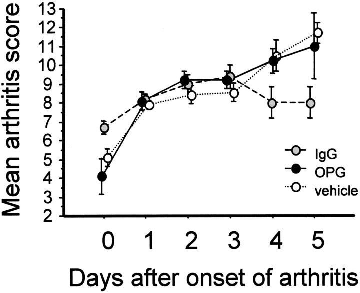
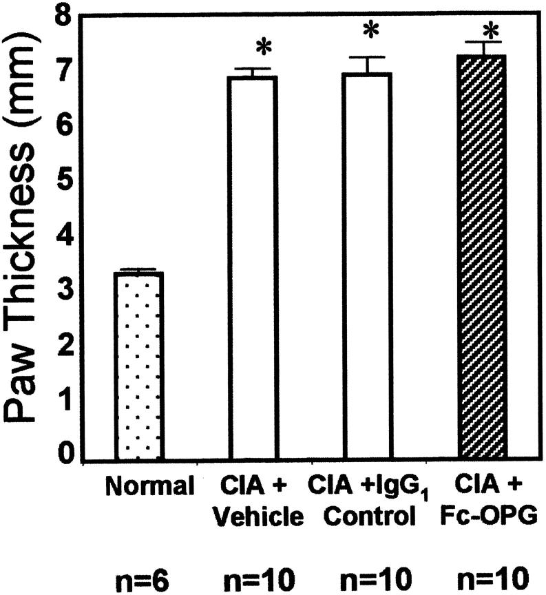
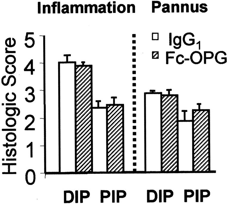
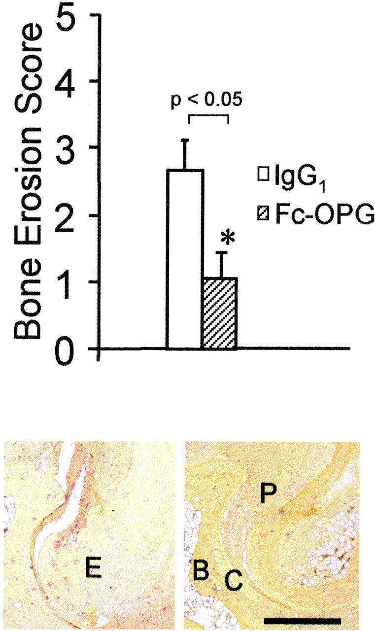
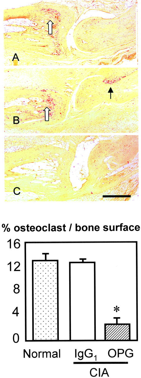
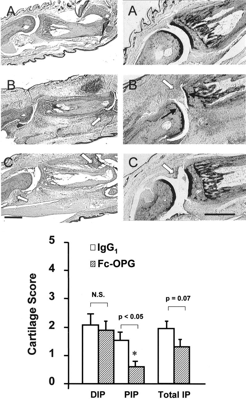
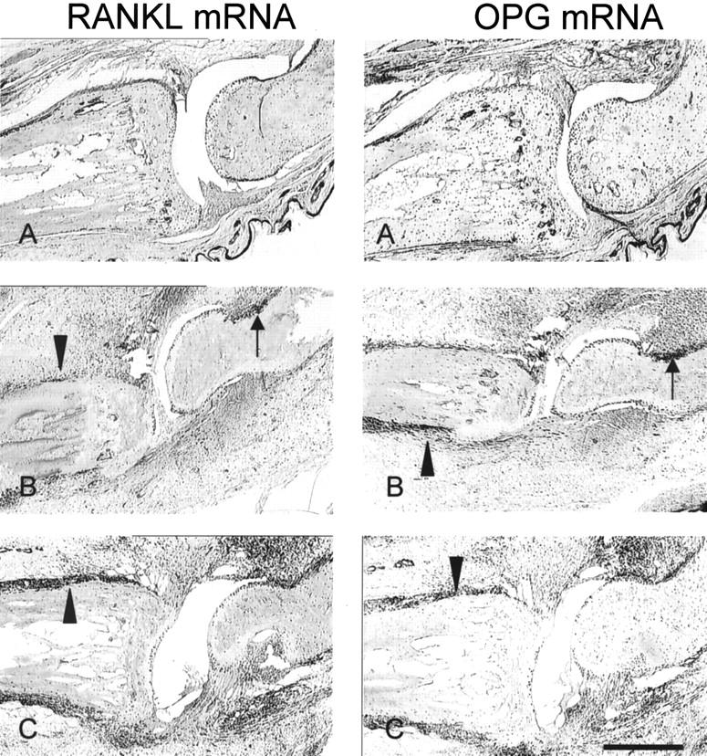
Rheumatoid arthritis is characterized by progressive synovial inflammation and joint destruction. While matrix metalloproteinases (MMPs) are implicated in the erosion of unmineralized cartilage, bone destruction involves osteoclasts, the specialized cells that resorb calcified bone matrix. RANK ligand (RANKL) expressed by stromal cells and T cells, and its cognate receptor, RANK, were identified as a critical ligand-receptor pair for osteoclast differentiation and survival. A decoy receptor for RANKL, osteoprotegerin, (OPG) impinges on this system and regulates osteoclast numbers and activity. RANKL is also expressed in collagen-induced arthritis (CIA) in which focal collections of osteoclasts are prominent at sites of bone destruction. To determine the role of RANK signaling events in the effector phase of CIA, we investigated effects of Fc-osteoprotegerin fusion protein (Fc-OPG) in CIA. After induction of CIA in Dark Agouti rats, test animals were treated with or without Fc-OPG (3 mg/kg/day) subcutaneously for 5 days, beginning at the onset of disease. Paraffin-embedded joints were then analyzed histologically and the adjacent bone assessed by histomorphometry. Osteoclasts were identified using TRAP staining and expression of the mRNA for OPG and RANKL was identified by in situ hybridization. The results indicated that short-term Fc-OPG effectively prevented joint destruction, even though it had no impact on the inflammatory aspects of CIA. In arthritic joints, Fc-OPG depleted osteoclast numbers by over 75% and diminished bone erosion scores by over 60%. Although cartilage loss was also reduced by Fc-OPG, the effects on cartilage were less striking than those on bone. In arthritic joints OPG mRNA was highly expressed and co-localized with RANK ligand, and treatment with Fc-OPG did not affect the expression of endogenous RANKL or OPG mRNA. These data demonstrate that short term Fc-OPG treatment has powerful anti-erosive effects, principally on bone, even though synovitis is not affected. These findings indicate the potential utility of disrupting RANK signaling to preserve skeletal integrity in inflammatory arthritis.


Figures

Figure 1.
Figure 1.
Kinetics of arthritis scores of CIA in DA rats. Rats were immunized with a single injection of CII and CIA developed in all animals 15 to 20 days later. At the onset of arthritis, they were randomly assigned to treatment groups for 5 days and arthritis was evaluated daily by a clinical score [as described in Ref. 29 ] (maximum score per animal, 16). Results show means (±SEM) for rats in each group.
Figure 2.
Figure 2.
Paw swelling in rats with CIA treated with and without Fc-OPG. The hind footpad thickness was assessed by caliper measurement at termination and bars represent the mean (±SEM) values for the animals in each group. (*, P < 0.01)
Figure 3.
Figure 3.
Histological evaluation of inflammation and pannus in CIA. The IP joints from arthritic rats (each group, n = 10) treated with human IgG1 or Fc-OPG were scored using a semi-quantitative system [as described in Materials and Methods]. Results are expressed as the mean scores (±SEM) for rats treated without or with OPG. DIP, distal IP joints; PIP, proximal IP joints.
Figure 4.
Figure 4.
Top: Effect of Fc-OPG on bone erosion scores. The IP joints from all arthritic rats treated without (n = 10) and with (n = 10) Fc-OPG were evaluated for bone erosions using a scoring system [as described in Materials and Methods]. Results are expressed as the mean scores (±SEM) of each group of animals (*, P < 0.05). Bottom: Osteoclasts at erosion sites in CIA (TRAP stain). Left: CIA + IgG1 control. Note TRAP-positive (red) cells eroding marginal and subchondral bone (E, erosion). Right: CIA + Fc-OPG. There is pannus (P) but absence of TRAP-positive osteoclasts or bone erosions. B, subchondral bone; C, cartilage. Bar, 500 μm.
Figure 5.
Figure 5.
Top: Effect of Fc-OPG on osteoclasts in CIA (TRAP-stain). Sagittal section of IP joint from normal rats (A), CIA rats treated with IgG1 (B) or CIA treated with Fc-OPG (C). Osteoclasts are colored red. Note osteoclasts in metaphysial bone (open arrow) and at focal bone erosion site (black arrow) in CIA controls (B) but not Fc-OPG treated CIA (C). Bar, 500 μm. Bottom: Effect of Fc-OPG on osteoclast surface. The percent osteoclast surface (Ocs/Bs) in the juxta-articular bone of non-arthritic and arthritic rats treated without or with Fc-OPG was determined. The bars represent the means (±SEM) for the animals in each group. (*, P < 0.01).
Figure 6.
Figure 6.
Top: Effect of Fc-OPG on cartilage. Consecutive sections show representative IP joints from normals (A), CIA treated with IgG1 (B) or CIA treated with Fc-OPG (C) for 5 days. Left: H&E stain. Right: Toluidine blue stains for cartilage. Note digital edema and synovial inflammation (arrowheads) in arthritic rats treated without or with Fc-OPG and cartilage loss (black arrows) in IgG1 controls. Bar, 500 μm. Bottom: Effect of Fc-OPG on cartilage erosion scores. The IP joints from arthritic rats treated without (n = 10) and with (n = 10) Fc-OPG were evaluated for cartilage erosions using a scoring system [as described in Materials and Methods]. Results are expressed as the mean (±SEM) scores for distal IP (DIP), proximal IP (PIP), or total IP joints of each intervention group. (*, P < 0.05).
Figure 7.
Figure 7.
In situ localization of RANKL and OPG mRNA in CIA. Consecutive IP joint sections were processed for DIG-labeled in situ hybridization as described in Methods. A, Normal; B, CIA + IgG1; C, CIA + Fc-OPG. Positive signal for the respective mRNAs is seen as dark purple staining. Note the high RANKL and OPG mRNA expression at the sites of bone erosion (black arrows) and periosteal inflammation (arrowheads). Bar, 500 μm.

References

    1. Goldring SR: The final pathogenetic steps in focal bone erosions in rheumatoid arthritis. Ann Rheum Dis 2000, 59(Suppl 1):i72-i74 - PMC - PubMed
    1. Gough A, Sambrook P, Devlin J, Huissoon A, Njeh C, Robbins S, Nguyen T, Emery P: Osteoclastic activation is the principal mechanism leading to secondary osteoporosis in rheumatoid arthritis. J Rheumatol 1998, 25:1282-1289 - PubMed
    1. Gravallese EM, Harada Y, Wang JT, Gorn AH, Thornhill TS, Goldring SR: Identification of cell types responsible for bone resorption in rheumatoid arthritis and juvenile rheumatoid arthritis. Am J Pathol 1998, 152:943-951 - PMC - PubMed
    1. Kong YY, Yoshida H, Sarosi I, Tan HL, Timms E, Capparelli C, Morony S, Oliveira-dos-Santos AJ, Van G, Itie A, Khoo W, Wakeham A, Dunstan CR, Lacey DL, Mak TW, Boyle WJ, Penninger JM: OPGL is a key regulator of osteoclastogenesis, lymphocyte development, and lymph-node organogenesis. Nature 1999, 397:315-323 - PubMed
    1. Lacey DL, Timms E, Tan HL, Kelley MJ, Dunstan CR, Burgess T, Elliott R, Colombero A, Elliott G, Scully S, Hsu H, Sullivan J, Hawkins N, Davy E, Capparelli C, Eli A, Qian YX, Kaufman S, Sarosi I, Shalhoub V, Senaldi G, Guo J, Delaney J, Boyle WJ: Osteoprotegerin ligand is a cytokine that regulates osteoclast differentiation and activation. Cell 1998, 93:165-176 - PubMed

Publication types

MeSH terms