. 2002 Oct 1;16(19):2530-43.
doi: 10.1101/gad.1024602.

c-Myc is essential for vasculogenesis and angiogenesis during development and tumor progression

Affiliations

c-Myc is essential for vasculogenesis and angiogenesis during development and tumor progression

Troy A Baudino et al. Genes Dev. .

Abstract

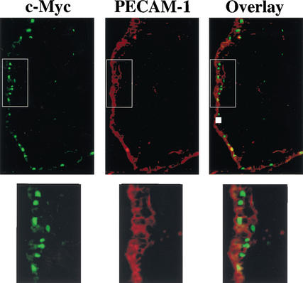
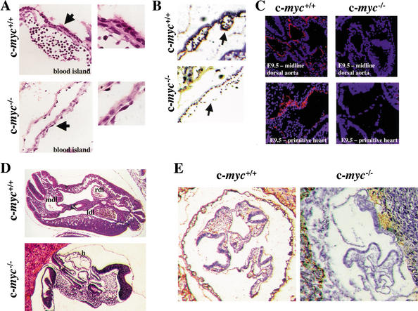
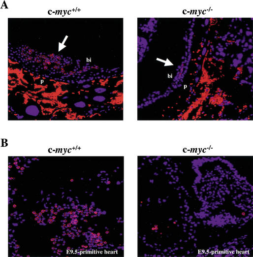
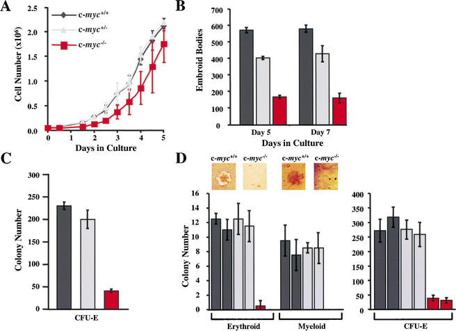
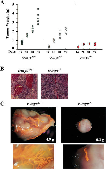
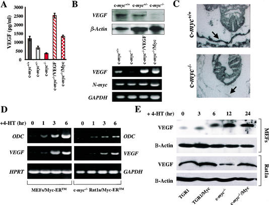
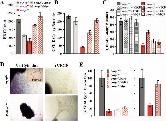
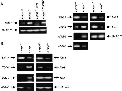
c-Myc promotes cell growth and transformation by ill-defined mechanisms. c-myc(-/-) mice die by embryonic day 10.5 (E10.5) with defects in growth and in cardiac and neural development. Here we report that the lethality of c-myc(-/-) embryos is also associated with profound defects in vasculogenesis and primitive erythropoiesis. Furthermore, c-myc(-/-) embryonic stem (ES) and yolk sac cells are compromised in their differentiative and growth potential. These defects are intrinsic to c-Myc, and are in part associated with a requirement for c-Myc for the expression of vascular endothelial growth factor (VEGF), as VEGF can partially rescue these defects. However, c-Myc is also required for the proper expression of other angiogenic factors in ES and yolk sac cells, including angiopoietin-2, and the angiogenic inhibitors thrombospondin-1 and angiopoietin-1. Finally, c-myc(-/-) ES cells are dramatically impaired in their ability to form tumors in immune-compromised mice, and the small tumors that sometimes develop are poorly vascularized. Therefore, c-Myc function is also necessary for the angiogenic switch that is indispensable for the progression and metastasis of tumors. These findings support the model wherein c-Myc promotes cell growth and transformation, as well as vascular and hematopoietic development, by functioning as a master regulator of angiogenic factors.


Figures

Figure 1
Figure 1
c-Myc is expressed in endothelial cells of the embryonic E8.5 yolk sac membrane. Endothelial cells were detected by immunofluorescence analyses with an antibody specific for PECAM-1 (red stain), which is expressed on the cell surface of endothelial cells. c-Myc expression was detected with a mouse-specific c-Myc antibody (green stain) and was largely confined to nuclei. Overlay shows that c-Myc-expressing cells (with green nuclei) also expressed PECAM-1. For clarity, the boxed areas in the top panels were magnified and are shown below.
Figure 2
Figure 2
c-myc−/− embryos have marked defects in vasculogenesis and angiogenesis. (A) Histological examination of H&E-stained yolk sacs of E8.5 wild-type versus c-myc−/− embryos. Arrows indicate area magnified to determine detailed morphology of cells lining the blood island (right panels). (B) IHC analyses of E9.5 yolk sacs from wild-type and c-myc−/− embryos with PECAM-1 antibody (brown stain). Arrows indicate blood islands. (C) Histological analyses of transverse sections of wild-type and c-myc−/− E9.5 embryos (top panels) and primitive hearts (bottom panels) with PECAM-1 antibody (red), counterstained with DAPI (blue). (D) H&E staining of wild-type and c-myc−/− embryos. (E) IHC analyses of E9.5 wild-type and c-myc−/− embryos with PECAM-1 antibody (brown stain).
Figure 3
Figure 3
c-myc−/− embryos have marked defects in primitive erythropoiesis. (A) Immunofluorescence analyses of transverse sections of E9.5 embryos using the erythroid-specific antibody Ter119 (red), counterstained with DAPI (blue). White arrows indicate blood islands. Bright red nonnucleated cells are maternal blood cells present in the placenta. (B) Immunofluorescence analyses of the cardiac region of E9.5 c-myc−/− and wild-type embryos using Ter-119 antibody and DAPI.
Figure 4
Figure 4
c-myc−/− ES cells are impaired in their in vitro differentiation. (A) Growth rates of wild-type (dark gray), c-myc+/− (light gray), and c-myc−/− (red) ES cells are shown in triplicate. (B) c-Myc deficiency impairs the primary differentiation of ES cells. The indicated ES cells were induced to undergo primary differentiation and after 5 or 7 d in culture, the numbers of EBs were determined. The results shown are the mean of three experiments performed in duplicate. (C) c-myc−/− ES cells are selectively impaired in secondary CFU-E colony formation. The indicated cells, wild-type (dark gray), c-myc+/− (light gray), and c-myc−/− (red), were plated in methylcellulose for 7 d. Then the EBs were dispersed and plated in methylcellulose containing Epo (CFU-E). Colony numbers were determined after 4 d. The data shown are representative of three experiments performed in duplicate. The mean number of colonies +/− the standard deviation is shown. (D) Hematopoietic progenitor colony assays were performed on E9.5 wild-type and c-myc−/− yolk sacs cultured in methylcellulose. Examples of erythroid-like and myeloid-like colonies formed by wild-type and c-myc−/− yolk sac cells in the absence of cytokines are shown above the left panel. In the right panel is shown CFU-E colony assays performed on individual E9.5 yolk sacs isolated from wild-type, c-myc+/−, and c-myc−/− yolk sacs, performed in duplicate.
Figure 5
Figure 5
c-myc−/− ES cells are impaired in tumorigenic and vasculogenic potential. (A) The weight of tumors arising in Scid mice following injection of wild-type (dark gray circles), c-myc+/− (light gray circles), or c-myc−/− (red circles) ES cells is shown. Tumors were harvested at the indicated intervals. (B) H&E staining of paraffin sections of tumors arising from c-myc−/− ES cells, compared with wild-type ES-cell-derived teratomas. (C) Dying Scid mice injected with wild-type or c-myc−/− ES cells were anesthetized and perfused with latex. The tumors were then resected and processed as described in Materials and Methods. Representative tumors are shown. The lower panels are magnified views showing capillary beds of the respective tumors.
Figure 6
Figure 6
c-Myc loss impairs VEGF expression. (A) VEGF production by wild-type (dark gray), c-myc+/− (light gray), and c-myc−/− (red) ES cells. The indicated cells were plated as described in Materials and Methods, and VEGF levels were determined by ELISA. c-myc−/− ES cells transduced with PGK–Myc (red checkerboard) or PGK–VEGF (red hatched) expression constructs were also analyzed for VEGF production. (B) Northern blot (top panel) and RT-PCR (bottom panel) analyses of VEGF transcripts in wild-type, c-myc+/−, and c-myc−/− ES cells, and in c-myc−/− ES cells transduced with the PGK–Myc or PGK–VEGF vectors (c-myc−/−/Myc and c-myc−/−/VEGF). Northern blots were hybridized with murine VEGF and actin probes. RT-PCR analyses of VEGF, N-myc, and GAPDH were determined. (C) Defects in VEGF expression are manifest in c-myc−/− embryos in vivo. In situ hybridization with a VEGF antisense probe on wild-type and c-myc−/− E8.5 embryo sections. Arrow indicates position of yolk sacs. Hybridization with the control sense VEGF probe failed to reveal signal (data not shown). (D) Myc activation is sufficient to induce VEGF expression. RT-PCR analyses of VEGF expression in primary MEFs and c-myc-null HO.15.19.3 Rat1a fibroblasts engineered to express a conditionally inducible form of c-Myc, Myc–ERTM, are shown. Cells were starved of serum overnight and then treated with 4-HT alone (1 μM). RNA was isolated at the indicated intervals, and RT-PCR analyses of VEGF, ODC, and GAPDH expression were performed. (E) VEGF protein levels are induced by c-Myc in primary and immortal fibroblasts. (Top panels) Levels of VEGF protein were determined by immunoblot analyses from primary MEFs engineered to express Myc–ERTM. Cells were treated with 4-HT for the indicated intervals, and VEGF protein levels were determined. β-Actin protein levels were determined as a loading control. (Bottom panels) VEGF protein levels were determined in wild-type (TGR1) and c-myc−/− (HO15.19.3) Rat1a-derived fibroblasts, and in these cells engineered to overexpress c-Myc.
Figure 7
Figure 7
VEGF rescues growth but not tumorigenic defects of c-myc−/− progenitors. (A) VEGF expression or the restoration of Myc rescues primary differentiation of c-myc−/− ES cells. The results shown are the mean of two experiments performed in quadruplicate. (B) VEGF expression or the restoration of c-Myc rescues secondary CFU-E colony formation of c-myc−/− EBs. The data shown are representative of three experiments performed in duplicate. The mean number of colonies +/− the standard deviation is shown. (C) The addition of exogenous VEGF, but not EGF or bFGF (all 10 ng/mL), rescues CFU-E colony formation of c-myc−/− EBs. Day 7 EBs were harvested and then plated in methylcellulose with Epo +/− the indicated cytokines. The data shown are representative of three experiments performed in duplicate. The mean number of colonies +/− the standard deviation is shown. (D) Yolk sac explant culture of E8.5 yolk sacs from wild-type (upper panels) or c-myc−/− (lower panels) embryos in serum-free medium (no cytokine) or in serum-free medium supplemented with 10 ng/mL VEGF (+VEGF). Photomicrographs were taken after 5 d of culture. (Inset at top left panel) Immunohistochemical staining of endothelial cells growing from these cultures with PECAM-1 antibody. (E) c-Myc, but not VEGF, expression rescues defects in tumor growth inherent to c-myc−/− ES cells. The weight of tumors arising in Scid mice following injection of 2 × 106 of the indicated ES cells is shown (wild-type, dark gray; c-myc−/−, red; c-myc−/−/puro, red speckled; c-myc−/−/VEGF, red hatched; c-myc−/−/Myc, red checkerboard). The frequency of teratoma formation was as follows: c-myc+/+, 100%; c-myc−/−, 50%; c-myc−/−/Myc, 80%; c-myc−/−/VEGF, 80%.
Figure 8
Figure 8
RT-PCR analyses of angiogenic and anti-angiogenic factors in c-myc−/− ES cells and c-myc−/− yolk sacs. (A, left) RT-PCR analyses of TSP-1 expression in wild-type, c-myc−/−, and c-myc−/− ES cells transduced with the PGK–Myc or PGK–VEGF vectors (c-myc−/−/Myc and c-myc−/−/VEGF). RNA was isolated from exponentially growing cells cultured in mLIF. (A, right) RT-PCR analyses of TSP-1, VEGF, ANG-1, ANG-2, Flk-1, Flt-1, and GAPDH expression in wild-type versus c-myc−/− ES cells. (B) RT-PCR analyses of ANG-1, ANG-2, Tie2, VEGF, TSP-1, Flk-1, Flt-1, and GAPDH expression in E9.5 wild-type and c-myc−/− yolk sacs.

References

    1. Adelman DM, Maltepe E, Simon MC. Multilineage embryonic hematopoiesis requires hypoxic ARNT activity. Genes & Dev. 1999;13:2478–2483. - PMC - PubMed
    1. Askew DS, Ashmun RA, Simmons BC, Cleveland JL. Constitutive c-myc expression in an IL-3-dependent myeloid cell line suppresses cell cycle arrest and accelerates apoptosis. Oncogene. 1991;6:1915–1922. - PubMed
    1. Barr LF, Campbell SE, Diette GB, Gabrielson EW, Kim S, Shim H, Dang CV. c-Myc suppresses the tumorigenicity of lung cancer cells and down-regulates vascular endothelial growth factor expression. Cancer Res. 2000;60:143–149. - PubMed
    1. Bello-Fernandez C, Packham G, Cleveland JL. Ornithine decarboxylase is a transcriptional target of c-myc. Proc Natl Acad Sci. 1993;90:7804–7808. - PMC - PubMed
    1. Blackwell TK, Huang J, Ma A, Kretzner L, Alt FW, Eisenman RN, Weintraub H. Binding of myc proteins to canonical and noncanonical DNA sequences. Mol Cell Biol. 1993;13:5216–5224. - PMC - PubMed

Publication types

MeSH terms

Substances