. 2002 Dec 20;277(51):49175-85.
doi: 10.1074/jbc.M205131200. Epub 2002 Oct 3.

Cloning and characterization of two extracellular heparin-degrading endosulfatases in mice and humans

Affiliations

Cloning and characterization of two extracellular heparin-degrading endosulfatases in mice and humans

Megumi Morimoto-Tomita et al. J Biol Chem. .

Abstract

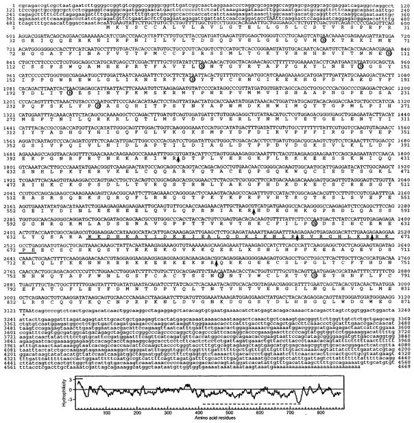
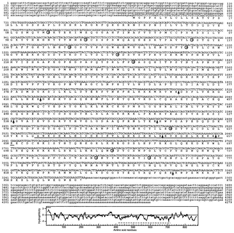
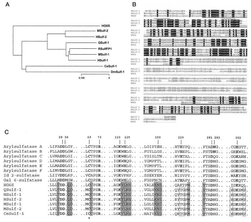
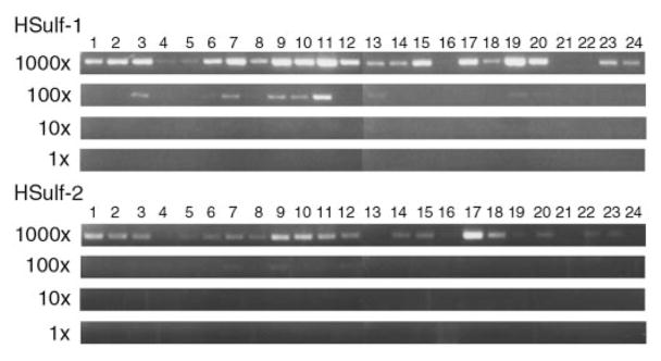
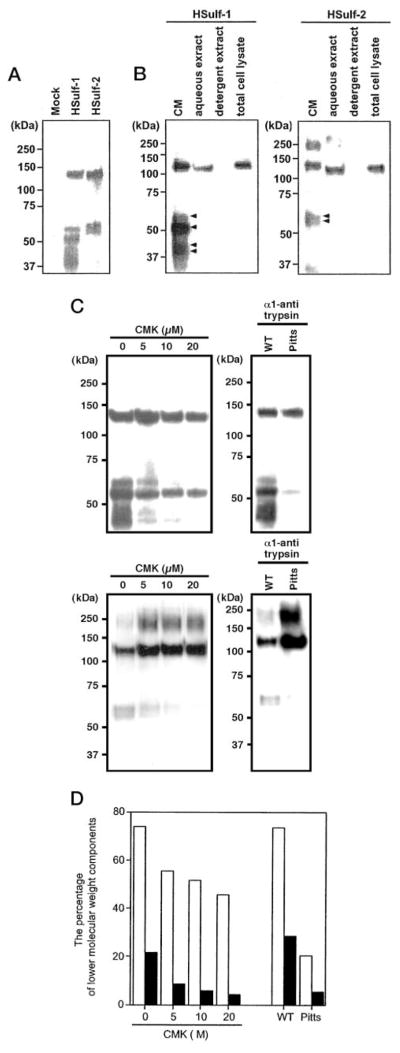
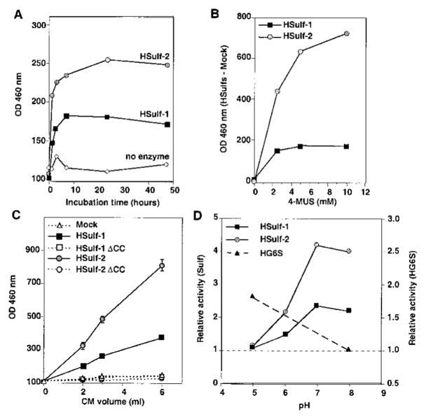
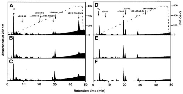
Here we report the cloning of a full-length cDNA encoding the human ortholog (HSulf-1) of the developmentally regulated putative sulfatases QSulf-1 (Dhoot, G. K., Gustafsson, M. K., Ai, X., Sun, W., Standiford, D. M., and Emerson, C. P., Jr. (2001) Science 293, 1663-1666) and RSulfFP1 (Ohto, T., Uchida, H., Yamazaki, H., Keino-Masu, K., Matsui, A., and Masu, M. (2002) Genes Cells 7, 173-185) as well as a cDNA encoding a closely related protein, designated HSulf-2. We have also obtained cDNAs for the mouse orthologs of both Sulfs. We demonstrate that the proteins encoded by both classes of cDNAs are endoproteolytically processed in the secretory pathway and are released into conditioned medium of transfected CHO cells. We demonstrate that the mammalian Sulfs exhibit arylsulfatase activity with a pH optimum in the neutral range; moreover, they can remove sulfate from the C-6 position of glucosamine within specific subregions of intact heparin. Taken together, our results establish that the mammalian Sulfs are extracellular endosulfatases with strong potential for modulating the interactions of heparan sulfate proteoglycans in the extracellular microenvironment.


Figures

Fig. 1
Fig. 1. HSulf-1 cDNA sequence and deduced amino acid sequence
Potential sites for N-linked glycosylation are indicated by circles. Potential furin cleavages sites are indicated by the arrows. The coiled-coil boundaries are underlined. Bottom panel, hydropathy analysis of the predicted protein. The line above the plot indicates the predicted signal sequence, and the line below is the hydrophilic region. The GenBank accession number is AY101175.
Fig. 2
Fig. 2. HSulf-2 cDNA sequence and deduced amino acid sequence
Potential sites for N-linked glycosylation are indicated by the circles. Potential furin cleavages sites are indicated by the arrows. The coiled-coil boundaries are underlined. Bottom panel, hydropathy analysis of the predicted protein. The line above the plot indicates the predicted signal sequence, and the line below is the hydrophilic region. The GenBank accession number is AY101176.
Fig. 3
Fig. 3. Genomic organization of Sulf genes
Long vertical bars indicate coding exons, whereas short vertical bars indicate noncoding exons. The box at the 3′-end denotes the last exon, which in all cases encodes the C-terminal 9 amino acids and also contains the sequence that is transcribed into the entire 3′-UTR of the mature cDNA. The intron-exon structures of the genes are described in greater detail in Table I. The sizes of the Sulf genes are ≥167 kb for HSulf-1 (gap within last intron after nucleotide 155,101 of the human Sulf-1 gene), 140.6 kb for MSulf-1, 129.2 kb for HSulf-2, and 81.7 kb for MSulf-2.
Fig. 4
Fig. 4. Sequence relationship among members of the Sulf family
A, dendrogram of the members of the Sulf family based on protein sequences. B, ClustalW alignment of HSulf-1, HSulf-2, and HG6S. Dark shading indicates identity for all three proteins, and gray shading signifies identity in two of the three proteins. The dark underline denotes the sulfatase domain. The short lines above the sequence indicate the two signature regions for sulfatases (PS00523 and PS00149) containing consensus sequences that are highly conserved among the entire sulfatase family. The star indicates the cysteine residue that is predicted to undergo an N-formylglycine modification. The dotted underline designates the hydrophilic region of the Sulfs. The double underline indicates the C-terminal region with homology among the three proteins. C, conservation of sulfatase domain active site residues within the Sulfs. Shown are Sulf sequences containing residues identical with (boldface letters) or homologous to the putative active sites of arylsulfatase A, numbered from its N terminus, based on the crystal structure (16). Gray boxes denote active site amino acids that are shared with HG6S, QSulf-1, HSulfs, MSulfs, and CeSulf-1, and open boxes denote residues that are shared within the Sulf family. The star indicates the cysteine residue that is predicted to undergo an N-formylglycine modification.
Fig. 5
Fig. 5. Detection of Sulf mRNAs in human tissues
cDNAs derived from various tissues were subjected to PCR to amplify products for HSulf-1 (top) or HSulf-2 (bottom). The cDNAs were diluted in 10-fold steps over a 1000-fold range. The tissues were as follows: 1, brain; 2, heart; 3, kidney; 4, spleen; 5, liver; 6, colon; 7, lung; 8, small intestine; 9, muscle; 10, stomach; 11, testis; 12, placenta; 13, salivary; 14, thyroid; 15, adrenal; 16, pancreas; 17, ovary; 18, uterus; 19, prostate; 20, skin; 21, plasma blood leukocytes; 22, bone marrow; 23, fetal brain; 24, fetal liver.
Fig. 6
Fig. 6. Expression of Sulf proteins in transfected CHO cells
A, conditioned medium of CHO cells, transfected with a cDNA for HSulf-1, HSulf-2, or the empty vector was collected, concentrated, and subjected to Western blotting for the Myc tag. B, the amount of Sulfs was determined by Western blotting in conditioned medium of cultured cells (CM), a high salt aqueous extract of crude membranes (aqueous extract), a detergent extract of the membrane residue remaining after the aqueous extraction (detergent extract), and a detergent extract of whole cells (total cell lysate). Equal numbers were used for each fraction. The arrows indicate the lower molecular weight species present in the CM. C, CHO cells were transfected with a cDNA for HSulf-1 (upper panel) or HSulf-2 in the presence of varying concentrations of CMK or were co-transfected with a cDNA encoding either wild-type α1-antitrypsin (WT) or the Pittsburgh mutant of α1-antitrypsin (active form of inhibitor, Pitts). The conditioned medium from each transfection was collected, concentrated, and subjected to Western blotting for detection of the Myc tag. The high molecular mass form of Sulf-2 (~250 kDa) that is observed in the presence of CMK or the active form of the antitrypsin inhibitor is presumed to be a SDS-resistant dimer. D, the bars indicate the percentage of lower molecular weight components (63, 58, 46, and 44 kDa for Hsulf-1 and 64 and 60 kDa for HSulf-2) relative to the total quantities of tagged proteins. The data were obtained by scanning the blots in C. Open bars show the results for HSulf-1, and solid bars show those for HSulf-2.
Fig. 7
Fig. 7. Arylsulfatase activity of expressed Sulfs and lack of the activity in HSulf mutants
HSulf-1, HSulf-2, or their mutated forms (HSulf-1 ΔCC and HSulf-2 ΔCC) were purified from the conditioned medium of transfected CHO cells by binding to Ni-NTA beads. A, the bead-bound material was tested for arylsulfatase activity as a function of time against 10 mM 4-MUS substrate at pH 8. The “no enzyme” control was based on testing conditioned medium from vector control-transfected CHO cells. No activity was detected in the absence of added substrate (not shown). The same results were obtained in three different experiments. B, the concentrated conditioned medium was tested for arylsulfatase activity at pH 8 for 2 h at different concentrations (1–10 mM) of 4-MUS. To eliminate background effects, the activity in vector control material was subtracted from that of Sulf-transfected material. C, the eluted material from Ni-NTA was tested for arylsulfatase activity at pH 8 for 2 h as a function of input volume of conditioned medium. The same results were obtained in three different experiments. D, bead-bound Sulfs were tested for arylsulfatase activity (1 h) at the indicated pH values. The activity of each Sulf was determined relative to that of beads exposed to an equivalent volume of vector-control conditioned medium. The activity of HG6S was determined (24-h incubation) relative to that of the buffer. The same results were obtained in three different experiments.
Fig. 8
Fig. 8. Endoglucosamine-6-sulfatase activity of expressed Sulfs
The conditioned medium of CHO cells transfected with the empty vector alone (A and D), HSulf-1 (B and E), or HSulf-2 (C and F) was prepared as described under “Experimental Procedures.” Porcine intestinal heparin (A–C) or shark cartilage chondroitin 6-sulfate (D–F) were incubated with the conditioned medium and then subsequently digested with either a mixture of bacterial heparinases or chondroitinase ABC. The resulting disaccharide fractions were analyzed by as described. The arrows correspond to the elution positions of authentic unsaturated disaccharide markers. The dotted lines indicate the concentrations of KH2PO4 used for elution.

References

    1. Parenti G, Meroni G, Ballabio A. Curr Opin Genet Dev. 1997;7:386–391. - PubMed
    1. Franco B, Meroni G, Parenti G, Levilliers J, Bernard L, Gebbia M, Cox L, Maroteaux P, Sheffield L, Rappold GA. Cell. 1995;81:15–25. - PubMed
    1. Puca AA, Zollo M, Repetto M, Andolfi G, Guffanti A, Simon G, Ballabio A, Franco B. Genomics. 1997;42:192–199. - PubMed
    1. Dhoot GK, Gustafsson MK, Ai X, Sun W, Standiford DM, Emerson CP., Jr Science. 2001;293:1663–1666. - PubMed
    1. Robertson DA, Freeman C, Morris CP, Hopwood JJ. Biochem J. 1992;288:539–544. - PMC - PubMed

Publication types

MeSH terms

Associated data