. 2002 Nov;76(21):10654-64.
doi: 10.1128/jvi.76.21.10654-10664.2002.

In situ localization and tissue distribution of the replication-associated proteins of Cucumber mosaic virus in tobacco and cucumber

Affiliations

In situ localization and tissue distribution of the replication-associated proteins of Cucumber mosaic virus in tobacco and cucumber

Fabrizio Cillo et al. J Virol. 2002 Nov.

Abstract

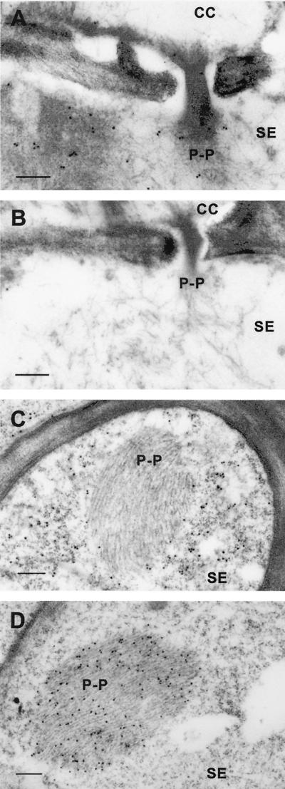
The replication-associated proteins encoded by Cucumber mosaic virus (CMV), the 1a and 2a proteins, were detected by immunogold labeling in two host species of this virus, tobacco (Nicotiana tabacum) and cucumber (Cucumis sativus). In both hosts, the 1a and 2a proteins colocalized predominantly to the vacuolar membranes, the tonoplast. While plus-strand CMV RNAs were found distributed throughout the cytoplasm by in situ hybridization, minus-strand CMV RNAs were barely detectable but were found associated with the tonoplast. In both cucumber and tobacco, 2a protein was detected at higher densities than 1a protein. The 1a and 2a proteins also showed quantitative differences with regard to tissue distributions in tobacco and cucumber. About three times as much 2a protein was detected in CMV-infected cucumber tissues as in CMV-infected tobacco tissues. In tobacco, high densities of these proteins were observed only in vascular bundle cells of minor veins. In contrast, in cucumber, high densities of 1a and 2a proteins were observed in mesophyll cells, followed by epidermis cells, with only low levels being observed in vascular bundle cells. Differences were also observed in the distributions of 2a protein and capsid protein in vascular bundle cells of the two host species. These observations may represent differences in the relative rates of tissue infection in different hosts or differences in the extent of virus replication in vascular tissues of different hosts.


Figures

FIG. 1.
FIG. 1.
In situ localization of 1a and 2a proteins by IGL in CMV-infected cucumber leaf cells. (A) Association of 1a protein with the tonoplast of the central vacuole (V) in a transection of a palisade mesophyll cell labeled with anti-1a protein antibody. c, chloroplast. The rectangle indicates the area enlarged in panels B and C. Scale bar, 500 nm. (B and C) Serial sections from the same cell as in panel A labeled with anti-1a protein and anti-2a protein antibodies, respectively, and showing colocalization of proteins 1a and 2a in association with the tonoplast. Scale bars, 250 nm. (D) Association of 1a protein with the tonoplast of a small vacuole (V) in a transection of a palisade mesophyll cell labeled with anti-1a protein antibody. The rectangle indicates the area enlarged in panels E and F. Scale bar, 250 nm. (E and F) Serial sections from the same cell as in panel D labeled with anti-1a protein and anti-2a protein antibodies, respectively, and showing colocalization of proteins 1a and 2a in association with the tonoplast. Scale bars, 100 nm. (G) Distribution of CP throughout the cytoplasm in a transection of an epidermis cell labeled with anti-CP antibody. V, vacuole. The rectangle indicates the area enlarged in panels H and I. Scale bar, 250 nm. (H and I) Serial sections from the same cell as in panel G labeled with anti-1a protein and anti-2a protein antibodies, respectively, and showing colocalization of proteins 1a and 2a in association with the tonoplast. Scale bars, 100 nm.
FIG.2.
FIG.2.
IGL of filamentous PP1 in tobacco leaf cells by anti-1a protein antibody. (A) Specific labeling by anti-1a protein antibody of PP1 (P-P) in a transection of a class III vein. PP1 aggregates spread through a pore in the sieve plate joining two SE. (B) Absence of PP1 labeling by anti-2a protein antibody in a different serial section of the same cells as in panel A. (C) Absence of PP1 labeling by anti-CP antibody in a transection of a minor (class IV) vein. The CP-specific signal was widespread throughout the cytoplasm of SE. (D) Specific labeling of PP1 by anti-1a protein antibody in a transection of a minor vein of a healthy plant leaf, showing that PP1 labeling was infection independent. Scale bars, 250 nm.
FIG. 3.
FIG. 3.
In situ localization of 1a and 2a proteins by IGL in a minor vein of a CMV-infected tobacco leaf. (A) Section of a class V immature vein of tobacco, used to identify the different cell types examined for the presence of CMV replication-associated proteins. BS, bundle sheath; X, xylem; XP, xylem parenchyma; PP, phloem parenchyma. The rectangle indicates the location of the area shown in panels B to D. Scale bar, 2 μm. (B to D) Serial sections from the same minor vein as in panel A labeled with anti-1a protein antibody (B) and anti-2a protein antibody (C and D). Proteins 1a and 2a colocalized in association with the tonoplast, and dense aggregates of gold particles were observed between individual vacuolar vesicles. Arrowheads indicate amorphous electron-dense areas specifically labeled by anti-2a protein antibody. Scale bars: B and D, 250 nm; C, 500 nm.
FIG. 4.
FIG. 4.
In situ localization of 2a protein by IGL in an immature SE of a CMV-infected leaf. (A) Localization of 2a protein to the tonoplast of vacuolar vesicles (V) in a transection of an SE in an immature minor vein of tobacco labeled with anti-2a protein antibody. PP, phloem parenchyma. Scale bar, 250 nm. (B) Localization of 2a protein to the tonoplast of a small vacuole (V) in a transection of an SE in an immature minor vein of cucumber labeled with anti-2a protein antibody. Scale bar, 100 nm.
FIG. 5.
FIG. 5.
Localization of viral RNA by ISH in vascular cells of veins of CMV-infected leaves. (A) Detection of CMV plus-strand RNA associated with paracrystalline arrays of virus particles (arrowheads) in a transection of a cucumber phloem parenchyma cell in a class IV or V vein. The plus-strand RNA was detected throughout the cytoplasm. In the inset, a larger aggregate of virions in a different phloem parenchyma cell is shown. Scale bars, 250 nm. (B) Detection of CMV plus-strand RNA in a transection of a tobacco CC of a minor vein. The CMV RNA-specific hybridization signal was widely dispersed in the cytoplasm, but clusters of gold particles were associated with the tonoplast around vacuolar vesicles (V). Scale bar, 250 nm. (C and D) Serial transections of a CMV-infected cucumber VB labeled with anti-2a protein antibody (C) and with a CMV minus-strand RNA-specific probe (D) and showing two small vacuoles, V1 and V2, in a phloem parenchyma cell. The 2a protein (C) and minus-strand RNA (arrowheads in D) were associated with the tonoplast of V1 and in between V1 and V2. Scale bars, 100 nm. The RNA probes were either complementary (A and B) or homologous (D) to the 3′-terminal 200 nt of CMV RNA 3.

References

    1. Buck, K. W. 1996. Comparison of the replication of positive-stranded RNA viruses of plants and animals. Adv. Virus Res. 47:159-251. - PMC - PubMed
    1. Canto, T., and P. Palukaitis. 1998. Transgenically expressed cucumber mosaic virus RNA 1 simultaneously complements replication of cucumber mosaic virus RNAs 2 and 3 and confers resistance to systemic infection. Virology 250:325-336. - PubMed
    1. Chen, J., and P. Ahlquist. 2000. Brome mosaic virus polymerase-like protein 2a is directed to the endoplasmic reticulum by helicase-like viral protein 1a. J. Virol. 74:4310-4318. - PMC - PubMed
    1. Clark, A. M., K. R. Jacobsen, D. E. Bostwick, J. M. Dannehoffer, M. I. Skaggs, and G. A. Thompson. 1997. Molecular characterization of a phloem-specific gene encoding the filament protein, phloem protein 1 (PP1), from Cucurbita maxima. Plant J. 12:49-61. - PubMed
    1. den Boon, J. A., J. Chen, and P. Ahlquist. 2001. Identification of sequences in brome mosaic virus replicase protein 1a that mediate association with endoplasmic reticulum membranes. J. Virol. 75:12370-12381. - PMC - PubMed

Publication types

LinkOut - more resources