Modulation of the cell division cycle by human papillomavirus type 18 E4
- PMID: 12368334
- PMCID: PMC136601
- DOI: 10.1128/jvi.76.21.10914-10920.2002
Modulation of the cell division cycle by human papillomavirus type 18 E4
Abstract
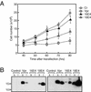
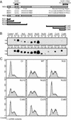
The life cycle of human papillomaviruses (HPVs) is tightly coupled to the differentiation program of their host epithelial cells. HPV E4 gene expression is first observed in the parabasal layers of squamous epithelia, suggesting that the E4 gene product contributes to the mechanism of differentiation-dependent virus replication, although its biological function remains unclear. We analyzed the effect of HPV type 18 E4 on cell proliferation and found that E4 expression induced cell cycle arrest at the G(2)/M boundary. The functional region of E4 necessary for the growth arrest activity was located in the central portion of the molecule, and this activity was independent of the E4-mediated collapse of cytokeratin intermediate filament structures.
Figures


Similar articles
-
Cooperation between different forms of the human papillomavirus type 1 E4 protein to block cell cycle progression and cellular DNA synthesis.J Virol. 2004 Dec;78(24):13920-33. doi: 10.1128/JVI.78.24.13920-13933.2004. J Virol. 2004. PMID: 15564500 Free PMC article.
-
Cutaneous and mucosal human papillomavirus E4 proteins form intermediate filament-like structures in epithelial cells.Virology. 1993 Nov;197(1):176-87. doi: 10.1006/viro.1993.1578. Virology. 1993. PMID: 8212552
-
Specific interaction between HPV-16 E1-E4 and cytokeratins results in collapse of the epithelial cell intermediate filament network.Nature. 1991 Aug 29;352(6338):824-7. doi: 10.1038/352824a0. Nature. 1991. PMID: 1715519
-
Role for Wee1 in inhibition of G2-to-M transition through the cooperation of distinct human papillomavirus type 1 E4 proteins.J Virol. 2006 Aug;80(15):7416-26. doi: 10.1128/JVI.00196-06. J Virol. 2006. PMID: 16840322 Free PMC article.
-
Novel Functions of the Human Papillomavirus E6 Oncoproteins.Annu Rev Virol. 2015 Nov;2(1):403-23. doi: 10.1146/annurev-virology-100114-055021. Epub 2015 Sep 2. Annu Rev Virol. 2015. PMID: 26958922 Review.
Cited by
-
Cooperation between different forms of the human papillomavirus type 1 E4 protein to block cell cycle progression and cellular DNA synthesis.J Virol. 2004 Dec;78(24):13920-33. doi: 10.1128/JVI.78.24.13920-13933.2004. J Virol. 2004. PMID: 15564500 Free PMC article.
-
Mutations in HPV18 E1^E4 Impact Virus Capsid Assembly, Infectivity Competence, and Maturation.Viruses. 2017 Dec 19;9(12):385. doi: 10.3390/v9120385. Viruses. 2017. PMID: 29257050 Free PMC article.
-
Condylomata acuminata: A retrospective analysis on clinical characteristics and treatment options.Heliyon. 2020 Mar 11;6(3):e03547. doi: 10.1016/j.heliyon.2020.e03547. eCollection 2020 Mar. Heliyon. 2020. PMID: 32190761 Free PMC article.
-
Mechanisms by which HPV Induces a Replication Competent Environment in Differentiating Keratinocytes.Viruses. 2017 Sep 19;9(9):261. doi: 10.3390/v9090261. Viruses. 2017. PMID: 28925973 Free PMC article. Review.
-
G2/M cell cycle arrest in the life cycle of viruses.Virology. 2007 Nov 25;368(2):219-26. doi: 10.1016/j.virol.2007.05.043. Epub 2007 Aug 6. Virology. 2007. PMID: 17675127 Free PMC article. Review.
References
-
- Andersen, B., A. Hariri, M. R. Pittelkow, and M. G. Rosenfeld. 1997. Characterization of Skn-1a/i POU domain factors and linkage to papillomavirus gene expression. J. Biol. Chem. 272:15905-15913. - PubMed
Publication types
MeSH terms
Substances
LinkOut - more resources
Full Text Sources

