. 2002 Nov;76(21):10942-50.
doi: 10.1128/jvi.76.21.10942-10950.2002.

A sensitive, quantitative assay for human immunodeficiency virus type 1 integration

Affiliations

A sensitive, quantitative assay for human immunodeficiency virus type 1 integration

Una O'Doherty et al. J Virol. 2002 Nov.

Abstract

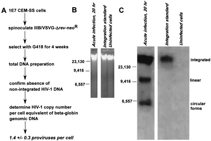
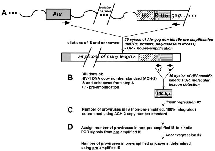
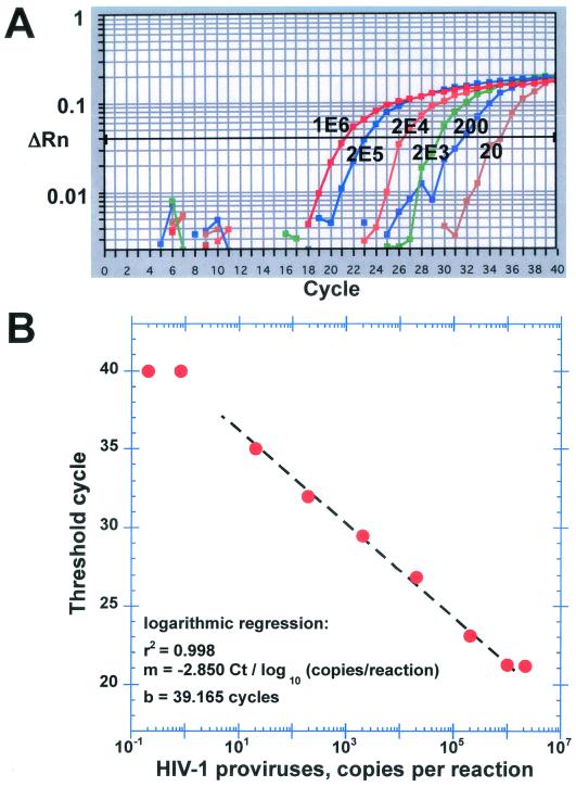
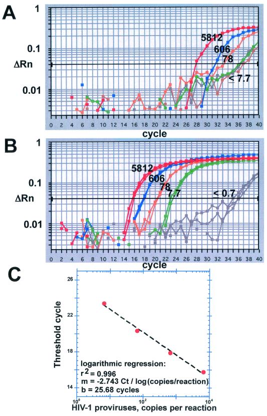
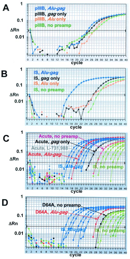
Quantitative methods to measure human immunodeficiency virus type 1 (HIV-1) integration promise to be important tools in dissecting the mechanisms whereby latent reservoirs of provirus are established, most notably in the resting T cells of patients receiving antiretroviral therapy. Here we describe a fluorescence-monitored, nested PCR assay that is able to quantify the relatively rare integration events that occur within these cells. Following DNA extraction, a nonkinetic preamplification step is performed with primers that bind genomic Alu elements and HIV-1 gag sequences, under conditions where primers, deoxynucleoside triphosphates, and enzyme are not limiting. This is followed by a kinetic PCR that quantitates HIV-1 long terminal repeat sequences. A T-cell-based integration standard which reflects the randomness of HIV-1 integration is also described. The assay is 10 to 100 times more sensitive than previously reported quantitative Alu PCR-based integration assays. It is specific for integration events, since no proviruses are detected in cells infected either in the presence of an integrase inhibitor or with an integrase-deficient virus. This method promises to provide important new insights into the processes underlying the accumulation and persistence of latent HIV-1 reservoirs and may eventually be useful clinically in monitoring the eradication of latent virus by novel therapies.


Figures

FIG. 1.
FIG. 1.
Preparation and characterization of the IS cell line. (A) Infection and selection strategy. (B) Equal amounts of DNA (20 μg/lane) from CEM-SS cells acutely infected with wild-type HIV-1IIIB (left lane), the antibiotic-selected IS line (center lane), or uninfected CEM-ss cells (right lane) were electrophoresed on a 0.8% agarose gel and stained with ethidium bromide prior to Southern blotting. (C) Autoradiogram of the Southern blot prepared from the gel in panel B. Whereas integrated, linear, and circular forms of HIV-1 DNA were present in the acutely infected cells (left lane), only integrated forms of HIV-1 DNA were present in the IS (center lane). Uninfected cells were free of HIV-1 signals (right lane). The intensities of the HIV-1 bands in the acutely infected sample (where DNA was prepared 20 h after viral exposure) are higher than those in the IS, because spinoculation delivered such a high multiplicity of infection to the CEM-SS-R5 cells that multiple proviruses were established in the majority of cells in the sample. In contrast, following 4 weeks of drug selection, cells in the IS contained a mean (± standard deviation) of 1.4 ± 0.3 proviruses per cell.
FIG. 2.
FIG. 2.
PCR and standardization strategy. (A) The nonkinetic PCR preamplification uses primers that bind genomic Alu and HIV-1 gag sequences. Substrates and enzyme are not limiting. Heterogeneous amplicons of variable length are produced. (B) Kinetic PCR to quantitate HIV-1-specific sequences within the LTR. (C) First regression, for provirus content of the IS. (D) Second regression, for provirus content of the unknowns.
FIG. 3.
FIG. 3.
The first regression, for provirus content of IS dilutions. An HIV-specific standard curve was generated using serial dilutions of genomic DNA from accurately counted ACH-2 cells. (A) Real-time monitoring of signals obtained from the ACH-2 standards. The known copy numbers in each dose of standard are shown over the corresponding amplification curves. 1E6 indicates 1,000,000 copies; 2E5 indicates 200,000 copies, etc. (B) Logarithmic regression to generate a line of best fit. Points outside the linear range are shown to illustrate the sigmoidal shape of the overall data set.
FIG. 4.
FIG. 4.
Second regression, for provirus content in unknowns. (A) The number of copies of HIV-1 DNA in dilutions of the nonpreamplified IS are shown over the corresponding amplification curves. These numbers were determined using the ACH-2 copy number standard (Fig. 3B). Since 100% of the HIV-1 DNA in the IS is integrated (Fig. 1), these numbers reflect the total number of proviruses in each reaction. (B) The number of proviruses per reaction in the nonpreamplified IS was assigned to the kinetic PCR signals from the preamplified IS, and a second standard line was constructed. These amplifications were performed in duplicate, with consistently close reproducibility. (C) Logarithmic regression of the preamplified IS signals. As few as eight proviruses per sample could be reliably measured in this run.
FIG.5.
FIG.5.
Sensitivity and specificity of the assay. (A) The plasmid pIIIB contains a full-length molecular clone of HIV-1IIIB and no human DNA (in particular, no Alu sequences). One-way preamplification with the Alu primer alone (orange) produces a kinetic PCR signal comparable to the signal from a matched dose of nonpreamplified plasmid (green). Signals roughly fivefold higher arise from either one-way preamplification with the gag primer alone (black) or bidirectional preamplification with both Alu and gag primers. (B) One-way preamplification of the IS using either the Alu primer alone (orange) or the gag primer alone (black) produces roughly a fivefold amplification relative to a matched sample of nonpreamplified IS (green). In contrast, the kinetic PCR signal obtained from the IS following bidirectional Alu-gag preamplification (blue) is shifted roughly 12 threshold cycles to the left, corresponding to a 4-log amplification over the nonpreamplified control. (C) Twelve hours after spinoculation of CEM-SS-R5 cells with HIV-1YU-2 (1,000 ng of p24Gag/ml), proviruses comprise only a minority of HIV-1 DNA. The integration signal from the acutely infected cells (red) falls within the linear range of the preamplified IS (blue curves, corresponding to 3,300, 1,000, 330, 100, 30, and 12 proviruses per reaction). One-way preamplification with the gag primer alone (black) generates a signal that is similar to the signal obtained when the infection is performed in the presence of a 10 μM dose of the integrase inhibitor L-731,988 (gray). This signal can be subtracted from the Alu-gag signal. It is produced via linear amplification of both integrated and unintegrated HIV-1 DNA. (D) Integration is not detected 12 h after spinoculation of CEM-SS-R5 cells with the HIV-1YU2-derived integrase-defective mutant virus D64A. Instead, Alu-gag preamplification results in a fivefold amplification, similar to one-way gag amplification.

References

    1. Bell, P., L. J. Montaner, and G. G. Maul. 2001. Accumulation and intranuclear distribution of unintegrated human immunodeficiency virus type 1 DNA. J. Virol. 75:7683-7691. - PMC - PubMed
    1. Benkirane, M., P. Corbeau, V. Housset, and C. Devaux. 1993. An antibody that binds the immunoglobulin CDR3-like region of the CD4 molecule inhibits provirus transcription in HIV-infected T cells. EMBO J. 12:4909-4921. - PMC - PubMed
    1. Bouyac-Bertoia, M., J. D. Dvorin, R. A. M. Fouchier, Y. Jenkins, B. E. Meyer, L. I. Wu, M. Emerman, and M. H. Malim. 2001. HIV-1 infection requires a functional integrase NLS. Mol. Cell 7:1025-1035. - PubMed
    1. Bukrinsky, M. I., T. L. Stanwick, M. P. Dempsey, and M. Stevenson. 1991. Quiescent T lymphocytes as an inducible virus reservoir in HIV-1 infection. Science 254:423-427. - PMC - PubMed
    1. Butler, S. L., M. S. T. Hansen, and F. D. Bushman. 2001. A quantitative assay for HIV DNA integration in vivo. Nat. Med. 7:631-634. - PubMed

Publication types

LinkOut - more resources