

Phosphorylation by the varicella-zoster virus ORF47 protein serine kinase determines whether endocytosed viral gE traffics to the trans-Golgi network or recycles to the cell membrane
- PMID: 12368341
- PMCID: PMC136633
- DOI: 10.1128/jvi.76.21.10980-10993.2002
Phosphorylation by the varicella-zoster virus ORF47 protein serine kinase determines whether endocytosed viral gE traffics to the trans-Golgi network or recycles to the cell membrane
Abstract
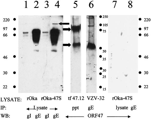
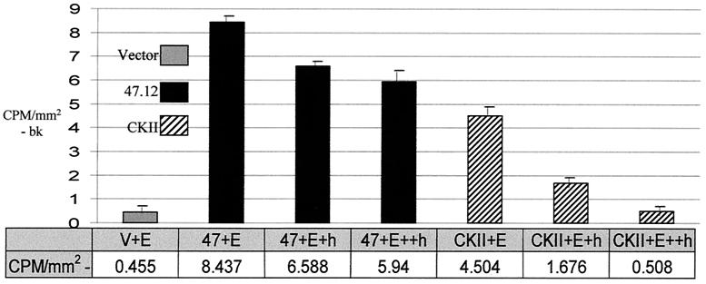
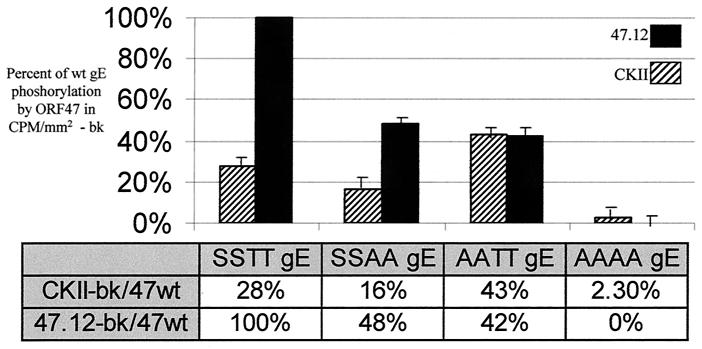
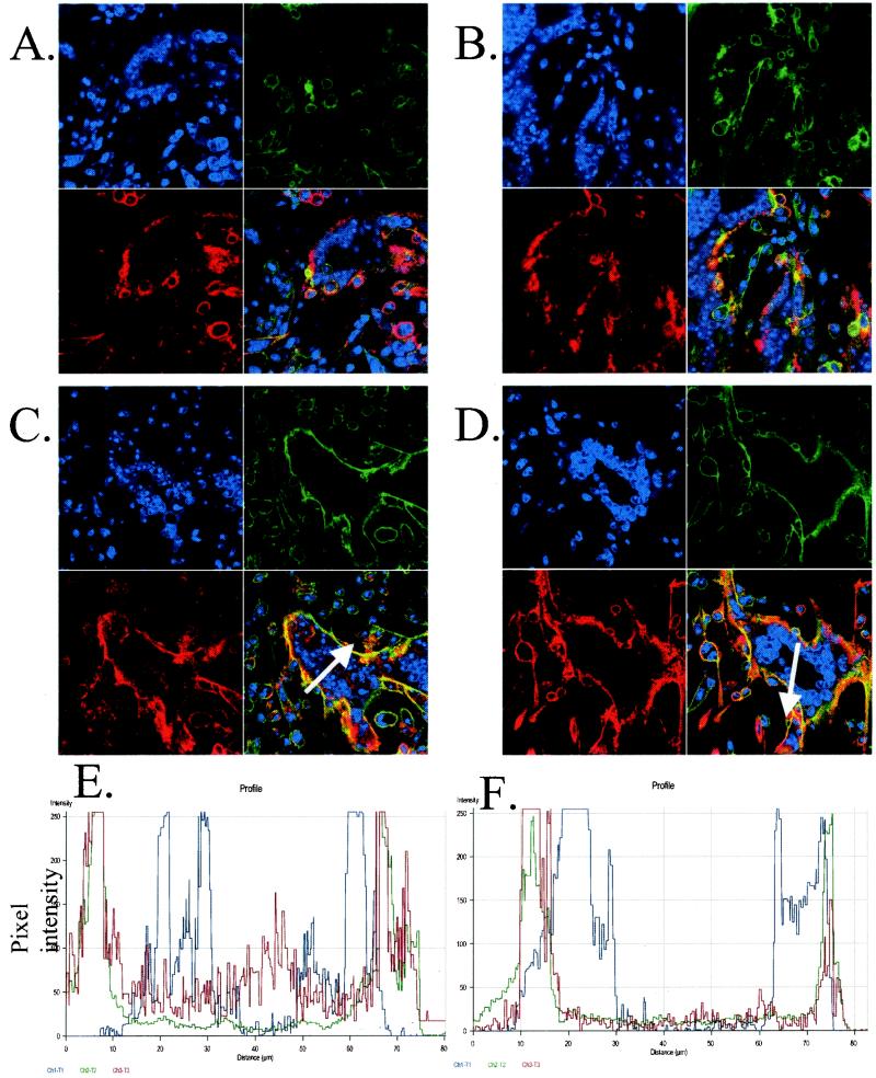
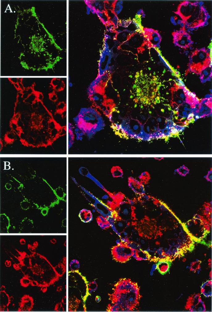
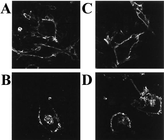
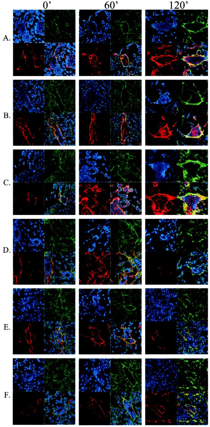
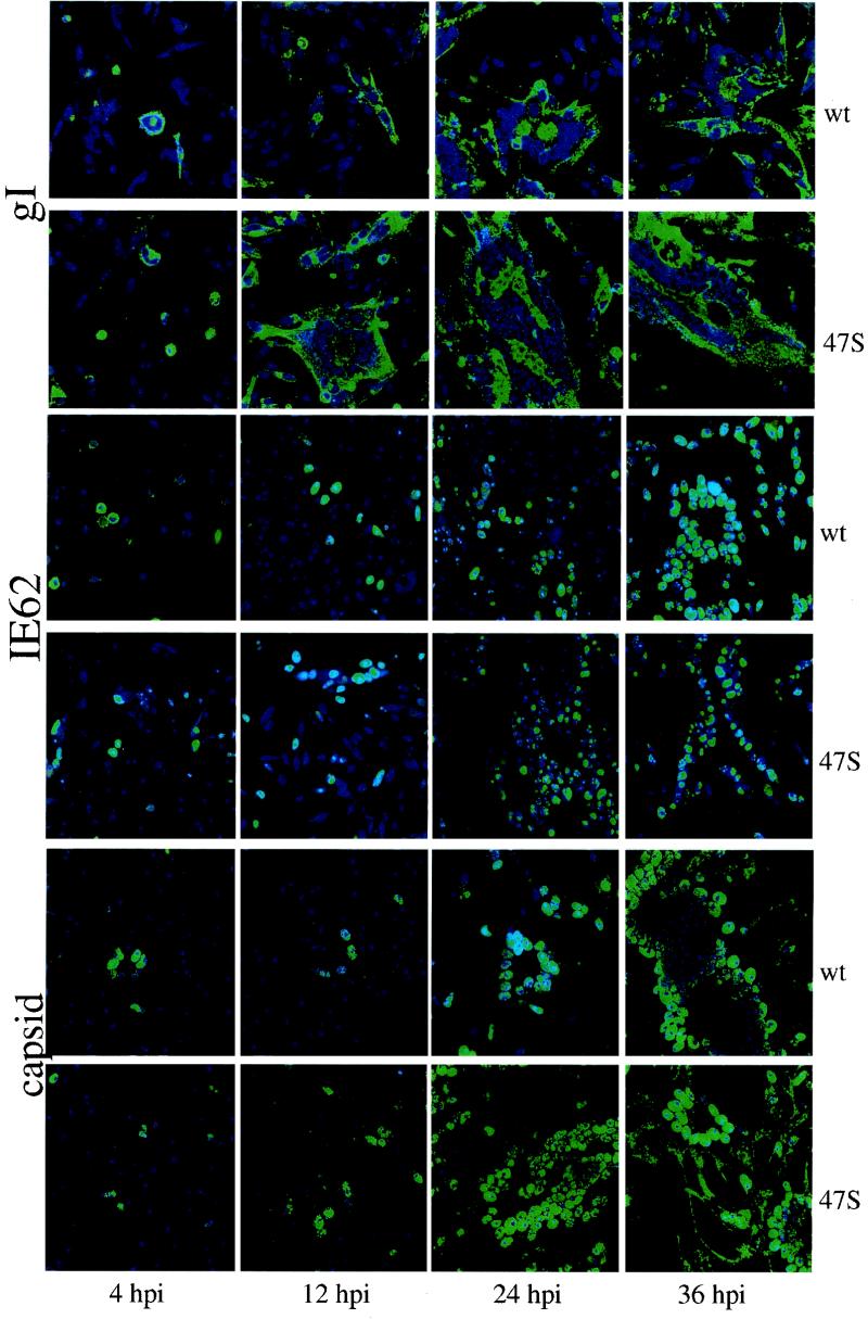
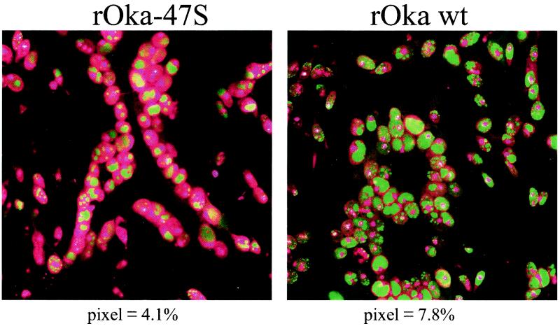
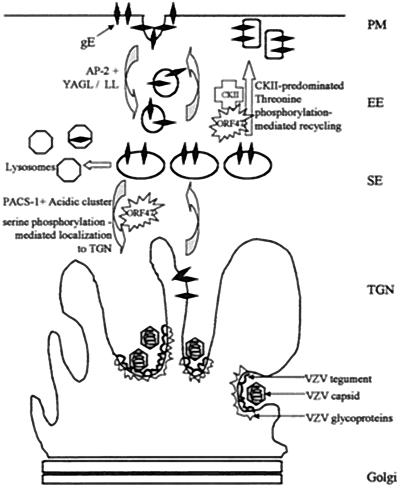
Like all alphaherpesviruses, varicella-zoster virus (VZV) infection proceeds by both cell-cell spread and virion production. Virions are enveloped within vacuoles located near the trans-Golgi network (TGN), while in cell-cell spread, surface glycoproteins fuse cells into syncytia. In this report, we delineate a potential role for serine/threonine phosphorylation of the cytoplasmic tail of the predominant VZV glycoprotein, gE, in these processes. The fact that VZV gE (formerly called gpI) is phosphorylated has been documented (E. A. Montalvo and C. Grose, Proc. Natl. Acad. Sci. USA 83:8967-8971, 1986), although respective roles of viral and cellular protein kinases have never been delineated. VZV ORF47 is a viral serine protein kinase that recognized a consensus sequence similar to that of casein kinase II (CKII). During open reading frame 47 (ORF47)-specific in vitro kinase assays, ORF47 phosphorylated four residues in the cytoplasmic tail of VZV gE (S593, S595, T596, and T598), thus modifying the known phosphofurin acidic cluster sorting protein 1 domain. CKII phosphorylated gE predominantly on the two threonine residues. In wild-type-virus-infected cells, where ORF47-mediated phosphorylation predominated, gE endocytosed and relocalized to the TGN. In cells infected with a VZV ORF47-null mutant, internalized VZV gE recycled to the plasma membrane and did not localize to the TGN. The mutant virus also formed larger syncytia than the wild-type virus, linking CKII-mediated gE phosphorylation with increased cell-cell spread. Thus, ORF47 and CKII behaved as "team players" in the phosphorylation of VZV gE. Taken together, the results showed that phosphorylation of VZV gE by ORF47 or CKII determined whether VZV infection proceeded toward a pathway likely involved with either virion production or cell-cell spread.
Figures
Similar articles
-
VZV ORF47 serine protein kinase and its viral substrates.Curr Top Microbiol Immunol. 2010;342:99-111. doi: 10.1007/82_2009_5. Curr Top Microbiol Immunol. 2010. PMID: 20186612 Review.
-
Comparison of varicella-zoster virus ORF47 protein kinase and casein kinase II and their substrates.J Med Virol. 2003;70 Suppl 1:S95-102. doi: 10.1002/jmv.10329. J Med Virol. 2003. PMID: 12627496
-
The varicella-zoster virus (VZV) open reading frame 47 (ORF47) protein kinase is dispensable for viral replication and is not required for phosphorylation of ORF63 protein, the VZV homolog of herpes simplex virus ICP22.J Virol. 1995 Nov;69(11):7367-70. doi: 10.1128/JVI.69.11.7367-7370.1995. J Virol. 1995. PMID: 7474171 Free PMC article.
-
Varicella-zoster virus (VZV) ORF65 virion protein is dispensable for replication in cell culture and is phosphorylated by casein kinase II, but not by the VZV protein kinases.Virology. 2001 Feb 1;280(1):62-71. doi: 10.1006/viro.2000.0741. Virology. 2001. PMID: 11162819
-
Glycoproteins encoded by varicella-zoster virus: biosynthesis, phosphorylation, and intracellular trafficking.Annu Rev Microbiol. 1990;44:59-80. doi: 10.1146/annurev.mi.44.100190.000423. Annu Rev Microbiol. 1990. PMID: 2174668 Review.
Cited by
-
Varicella-zoster virus infection of human foreskin fibroblast cells results in atypical cyclin expression and cyclin-dependent kinase activity.J Virol. 2006 Jun;80(11):5577-87. doi: 10.1128/JVI.00163-06. J Virol. 2006. PMID: 16699039 Free PMC article.
-
Deletion of the first cysteine-rich region of the varicella-zoster virus glycoprotein E ectodomain abolishes the gE and gI interaction and differentially affects cell-cell spread and viral entry.J Virol. 2009 Jan;83(1):228-40. doi: 10.1128/JVI.00913-08. Epub 2008 Oct 22. J Virol. 2009. PMID: 18945783 Free PMC article.
-
Roscovitine, a cyclin-dependent kinase inhibitor, prevents replication of varicella-zoster virus.J Virol. 2004 Mar;78(6):2853-62. doi: 10.1128/jvi.78.6.2853-2862.2004. J Virol. 2004. PMID: 14990704 Free PMC article.
-
Functions of the C-terminal domain of varicella-zoster virus glycoprotein E in viral replication in vitro and skin and T-cell tropism in vivo.J Virol. 2004 Nov;78(22):12406-15. doi: 10.1128/JVI.78.22.12406-12415.2004. J Virol. 2004. PMID: 15507627 Free PMC article.
-
Differentiation of varicella-zoster virus ORF47 protein kinase and IE62 protein binding domains and their contributions to replication in human skin xenografts in the SCID-hu mouse.J Virol. 2003 May;77(10):5964-74. doi: 10.1128/jvi.77.10.5964-5974.2003. J Virol. 2003. PMID: 12719588 Free PMC article.
References
Publication types
MeSH terms
Substances
Grants and funding
LinkOut - more resources
Full Text Sources
Other Literature Sources
Research Materials
Miscellaneous
