. 2002 Nov;76(21):10994-1002.
doi: 10.1128/jvi.76.21.10994-11002.2002.

A new type of adenovirus vector that utilizes homologous recombination to achieve tumor-specific replication

Affiliations

A new type of adenovirus vector that utilizes homologous recombination to achieve tumor-specific replication

Kathrin Bernt et al. J Virol. 2002 Nov.

Abstract

We have developed a new class of adenovirus vectors that selectively replicate in tumor cells. The vector design is based on our recent observation that a variety of human tumor cell lines support DNA replication of adenovirus vectors with deletions of the E1A and E1B genes, whereas primary human cells or mouse liver cells in vivo do not. On the basis of this tumor-selective replication, we developed an adenovirus system that utilizes homologous recombination between inverted repeats to mediate precise rearrangements within the viral genome resulting in replication-dependent activation of transgene expression in tumors (Ad.IR vectors). Here, we used this system to achieve tumor-specific expression of adenoviral wild-type E1A in order to enhance viral DNA replication and spread within tumor metastases. In vitro DNA replication and cytotoxicity studies demonstrated that the mechanism of E1A-enhanced replication of Ad.IR-E1A vectors is efficiently and specifically activated in tumor cells, but not in nontransformed human cells. Systemic application of the Ad.IR-E1A vector into animals with liver metastases achieved transgene expression exclusively in tumors. The number of transgene-expressing tumor cells within metastases increased over time, indicating viral spread. Furthermore, the Ad.IR-E1A vector demonstrated antitumor efficacy in subcutaneous and metastatic models. These new Ad.IR-E1A vectors combine elements that allow for tumor-specific transgene expression, efficient viral replication, and spread in liver metastases after systemic vector application.


Figures

FIG. 1.
FIG. 1.
Replication-dependent activation of E1A expression with positive-feedback mechanism. (A) Structures of Ad.AP, Ad.IR-AP, and Ad.IR-AP/E1A vectors. In Ad.AP, the AP reporter gene is under the direct control of the RSV long terminal repeat promoter. In Ad.IR-AP, the AP gene sequence is in the 3′→5′ orientation relative to the RSV promoter and flanked by two homology elements in inverse orientation (IR). As IRs, two rabbit β-globin introns that do not contain any transcription stop sites and are spliced out upon transcription were used. A bidirectional simian virus 40 polyadenylation signal (pA) was used to terminate transcription of the AP or E1A genes and to prevent the formation of antisense RNA from the promoter 3′ of the cassette. (B) Schematic of E1A-enhanced Ad.IR replication. During replication in tumor cells, homologous recombination between thetwo inverted repeats mediates the formation of a delta product carrying two invested copies of the left half of the parental genome(including the RSV promoter), flanking the transgene. In the delta product, the AP transgene is in the correct (5′→3′) orientation toward the RSV promoter. The structure of Ad.IR-AP/E1A is similar to that of Ad.IR-AP but allows bicistronic expression of AP together with E1A from the delta product. E1A expression subsequently enhances viral replication of the parental vectors, which, in turn, leads to more recombination events and a further increase in E1A expression. (C) AP expression in HeLa and SAEC after infection with Ad.AP, Ad.IR-AP, and Ad.IR-AP/E1A. HeLa cells and SAEC were infected with virus at an MOI of 30 PFU/cell for 3 h. After 48 h, cells were subjected to AP staining.
FIG. 2.
FIG. 2.
DNA replication kinetics of Ad.IR-AP, Ad.IR-AP/E1A, and Ad.WT in HeLa cells. A methylated input virus (8-kb fragment) can be distinguished from replicated (demethylated) progeny virus (6.5-kb fragment) by Southern blot analysis. At different time points after infection with Ad.IR-AP, Ad.IR-AP/E1A, and Ad.WT at an MOI of 100 PFU/cell, the amount of replicated genomes was determined in HeLa cells. There was no detectable Ad.WT replication at 6 h postinfection (data not shown).
FIG. 3.
FIG. 3.
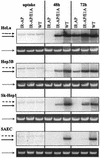
Viral DNA replication in different tumor cell lines and SAEC. HeLa, Hep3B, SK-Hep1, and SAEC cells were infected with Ad.IR-AP (IR-AP), Ad.IR-AP/E1A (IR-AP/E1A), and Ad.WT (WT) at an MOI of 30 PFU/cell for 3 h. To demonstrate similar uptake efficiencies, a set of cells was harvested 3 h after the start of infection (uptake). To analyze vector DNA replication, cells were harvested 48 and 72 h after infection (replication). The amount of input (8-kb [dashed arrow]) and replicated (6.5-kb [solid arrow]) genomes was determined by Southern blotting. The corresponding ethidium bromide-stained agarose gel is shown below each blot to demonstrate equal loading (chromosomal DNA [dotted arrow]). Blots with uptake lanes were exposed twice as long as the other blots. The comparable infectivities of the different cell lines are reflected by the similar intensities of the uptake lanes.
FIG. 4.
FIG. 4.
CPE after viral infection. HeLa, Hep3B, SK-Hep1, and SAEC cells were infected with Ad.IR-AP, Ad.IR-AP/E1A, and Ad.WT at MOIs ranging from 0.001 to 100 PFU/cell. After 7 days, cells were stained with crystal violet.
FIG. 5.
FIG. 5.
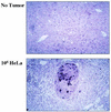
Tumor specificity of AP expression from Ad.IR-AP/E1A. CB17 mice were injected with 2 × 10⁶ HeLa cells via the portal vein to establish liver metastases. Fourteen days after transplantation of the cells, mice received 5 × 10⁹ PFU of Ad.IR-AP/E1A via tail vein injection. Six days after virus injection, mice were sacrificed, and liver sections were stained for AP expression. The lower panel shows a representative metastasis. The top panel shows a liver section of a naive mouse (without transplanted HeLa cells) that received i.v. Ad.IR-AP/E1A injection.
FIG. 6.
FIG. 6.
Spread of Ad.IR-AP/E1A through metastases over time. CB17 mice were injected with 2 × 10⁶ HeLa cells via the portal vein to establish liver metastases. Fourteen days after transplantation of the cells, groups of six mice received 5 × 10⁹ PFU of either Ad.IR-AP or Ad.IR-AP/E1A via tail vein injection. At 2, 5, and 10 days after virus injection, mice were sacrificed, and liver sections were stained for AP expression. Representative metastases are shown.
FIG. 7.
FIG. 7.
Inhibition of tumor growth depending on the number of initially transduced cells. HeLa cells were infected with Ad.IR-AP or Ad.IR-AP/E1A for 3 h at an MOI of 100 PFU/cell. Infected cells were trypsinized, washed (to remove noninternalized virus), and mixed with noninfected cells at different ratios (0.1, 1, and 10% infected cells). The cell mixture was injected into the inguinal region of immunodeficient mice. The tumor size was measured every other day. Time (t) is shown on the x axis. Individual data points represent the average volumes of four tumors; error bars were omitted for clarity. Statistical significance is indicated as follows: , statistically significant (P < 0.05) mean tumor size at day 25 compared to the uninfected control; ∗∗, statistically significant (P < 0.05) delay in tumor growth compared to the uninfected control; () and (∗∗), borderline statistical significance (P < 0.1).
FIG. 8.
FIG. 8.
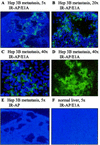
Replication of Ad.IR-AP/E1A in Hep3B metastases. CB17 mice received 2 × 10⁶ Hep3B cells via the portal vein to establish liver metastases (A to E) or buffer (F). Fourteen days after transplantation of the cells, groups of three mice were injected via the tail vein with 5 × 10⁹ PFU of either Ad.IR-AP/E1A (A to D and F) or Ad.IR-AP (E) on two consecutive days (total dose of 1010 PFU). For histological analyses, mice were sacrificed 10 days after the last virus injection. Frozen liver sections were stained for viral hexon and detected with a secondary fluorescein isothiocyanate-conjugated antibody (green) (A to F). Cell nuclei were counterstained with DAPI (blue) (A to C, E, and F). Representative sections are shown. Note that at the time of analysis, only de novo-expressed hexon, not the capsid hexon present in incoming particles, is detected. The magnification of each section is indicated.
FIG. 9.
FIG. 9.
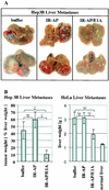
Antitumor effect of Ad.IR-AP/E1A in two models for liver metastasis. CB17 mice received 2 ×10⁶ Hep3B or HeLa cells via the portal vein to establish liver metastases. Fourteen days after transplantation of the cells, groups of four mice were injected via the tail vein with 5 × 10⁹ PFU of either Ad.IR-AP/E1A or Ad.IR-AP or with virus dilution buffer (buffer) on two consecutive days (total dose of 1010 PFU). When the control mice started to exhibit signs of ascites (4 and 9 weeks after transplantation of HeLa and Hep3B cells, respectively), all mice were sacrificed. (A) Hep3B-derived liver metastases. Macroscopic aspects of livers bearing the smallest (upper) and largest (lower) metastases in each group. (B) Analysis of Hep3B- and HeLa-derived metastases. Tumor tissue derived from Hep3B cells was isolated from liver tissue by microdissection under a stereomicroscope. The total liver weight and tumor weights were measured. The tumor burden was expressed as a percentage of tumor weight of the total liver weight (left panel). Microdissection of the HeLa tumor was technically impossible. Therefore, the total weight of tumor-bearing livers was determined as an estimate of tumor burden (right panel). Statistical significance is indicated as follows: , statistically significant (P < 0.05); ∗∗, borderline statistically significant (P < 0.1); ns, not statistically significant.

References

    1. Alemany, R., C. Balague, and D. T. Curiel. 2000. Replicative adenoviruses for cancer therapy. Nat. Biotechnol. 18:723-727. - PubMed
    1. Babiss, L. E., J. M. Friedman, and J. E. Darnel, Jr. 1986. Cellular promoter incorporated into adenovirus genome: effects of viral regulatory elements on transcription rates and cell specificity of albumin and beta-globin promoters. Mol. Cell. Biol. 6:3798-3806. - PMC - PubMed
    1. Bernt, K. M., D. S. Steinwaerder, S. Ni, Z.-Y. Li, S. R. Roffler, and A. Lieber. 2002. Enzyme-activated prodrug therapy enhances tumor-specific replication of adenovirus vectors. Cancer Res., in press. - PubMed
    1. Bischoff, J. R., D. H. Kirn, A. Williams, C. Heise, S. Horn, M. Muna, L. Ng, J. A. Nye, A. Sampson-Johannes, A. Fattaey, and F. McCormick. 1996. An adenovirus mutant that replicates selectively in p53-deficient human tumor cells. Science 274:373-376. - PubMed
    1. Bruder, J. T., A. Appiah, W. M. Kirkman III, P. Chen, J. Tian, D. Reddy, D. E. Brough, A. Lizonova, and I. Kovesdi. 2000. Improved production of adenovirus vectors expressing apoptotic transgenes. Hum. Gene Ther. 11:139-149. - PubMed

Publication types

MeSH terms

LinkOut - more resources