. 2002 Nov;76(21):11091-103.
doi: 10.1128/jvi.76.21.11091-11103.2002.

Synergistic activation of human immunodeficiency virus type 1 promoter activity by NF-kappaB and inhibitors of deacetylases: potential perspectives for the development of therapeutic strategies

Affiliations

Synergistic activation of human immunodeficiency virus type 1 promoter activity by NF-kappaB and inhibitors of deacetylases: potential perspectives for the development of therapeutic strategies

Vincent Quivy et al. J Virol. 2002 Nov.

Abstract

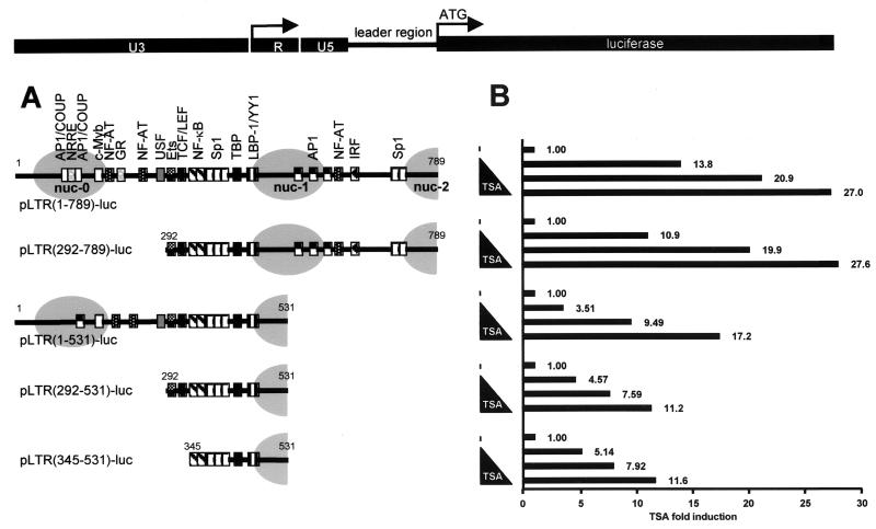
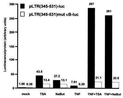
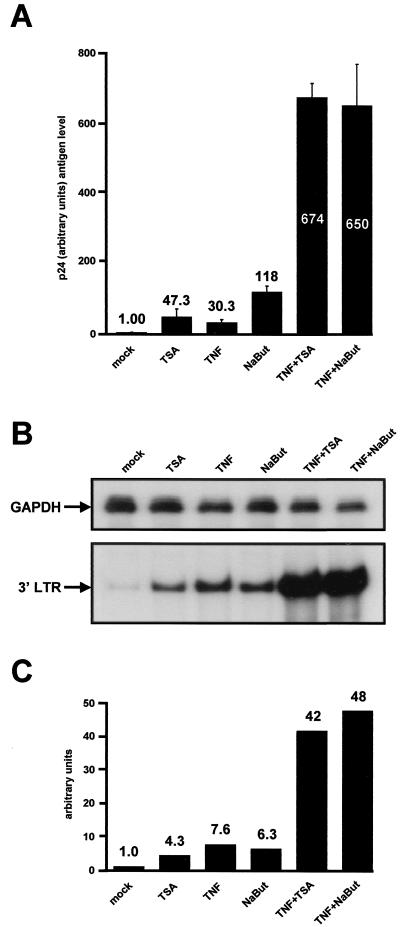
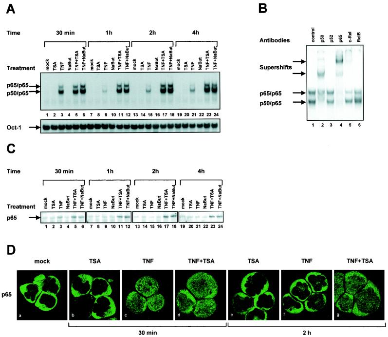
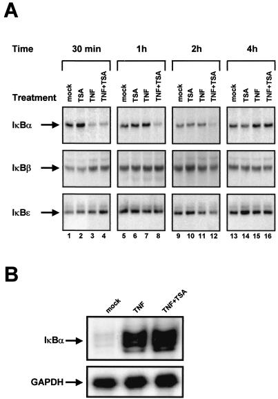
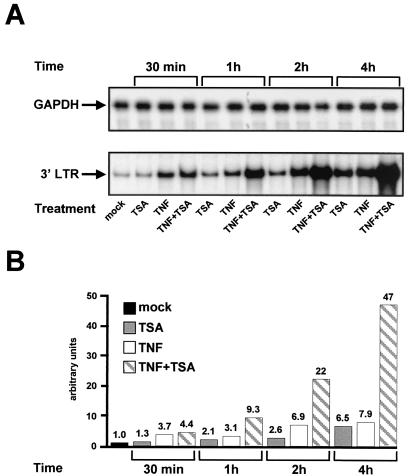
The transcription factor NF-kappaB plays a central role in the human immunodeficiency virus type 1 (HIV-1) activation pathway. HIV-1 transcription is also regulated by protein acetylation, since treatment with deacetylase inhibitors such as trichostatin A (TSA) or sodium butyrate (NaBut) markedly induces HIV-1 transcriptional activity of the long terminal repeat (LTR) promoter. Here, we demonstrate that TSA (NaBut) synergized with both ectopically expressed p50/p65 and tumor necrosis factor alpha/SF2 (TNF)-induced NF-kappaB to activate the LTR. This was confirmed for LTRs from subtypes A through G of the HIV-1 major group, with a positive correlation between the number of kappaB sites present in the LTRs and the amplitude of the TNF-TSA synergism. Mechanistically, TSA (NaBut) delayed the cytoplasmic recovery of the inhibitory protein IkappaBalpha. This coincided with a prolonged intranuclear presence and DNA binding activity of NF-kappaB. The physiological relevance of the TNF-TSA (NaBut) synergism was shown on HIV-1 replication in both acutely and latently HIV-infected cell lines. Therefore, our results open new therapeutic strategies aimed at decreasing or eliminating the pool of latently HIV-infected reservoirs by forcing viral expression.


Figures

FIG. 1.
FIG. 1.
TSA responsiveness of different HIV-1 LTRs with deletions. (A) Organization of the 5′ LTR region of the prototype HIV-1 virus LAI (subtype B). The LTR is composed of the U3, R, and U5 regions. The complete LTR and the leader region (nt 1 to 789) (nt +1 is the start of U3 in the 5′ LTR) control the luciferase reporter gene. The transcription factor binding sites present in the different LTRs with deletions are detailed for each construct and are aligned with nucleosome positioning (grey ovals). The following transcription factor binding sites are present in the full-length LTR construct (reviewed in reference 38): two AP1/COUP sites, the nuclear receptor-responsive element (NRRE), one c-Myb site, three NF-AT sites, one GR site, one USF site, one Ets-1 site, one TCF/LEF site, two κB sites, five Sp1 sites, the TATA box, one LBP-1/YY1 site, three AP-1 sites, and one IRF site. (B) SupT1 cells were transiently transfected with 500 ng of each reporter construct containing the different HIV-1 LTRs with deletions, and cells were treated with increasing concentrations of TSA. Luciferase activities were measured in cell lysates. To compare the TSA responsiveness of the different LTRs, basal LTR activity of each construct was arbitrarily set at a value of 1. Results are presented as histograms indicating the TSA inductions of the constructs with respect to their respective basal activity. Values represent the means of duplicate samples. An experiment representative of three independent transfections is shown. Variation for a given plasmid between different experiments was <15% in most cases.
FIG. 2.
FIG. 2.
Synergistic activation of HIV-1 promoter by cytokine TNF and inhibitors of deacetylases, TSA, and NaBut. SupT1 cells were transiently transfected with 500 ng of the indicated constructs. Cells were mock treated or treated with TSA, NaBut, TNF, TNF-TSA, or TNF-NaBut. The mock-treated value of the wild-type LTR reporter construct was arbitrarily set at a value of 1. Values represent the means of duplicate samples. A representative experiment of three independent transfections is shown. Variation for a given plasmid between different experiments was <15% in most cases.
FIG. 3.
FIG. 3.
Synergistic activation by TSA and TNF of the LTRs from the HIV-1 group M subtypes A through G. SupT1 cells were transiently transfected with 500 ng of an LTR luciferase construct containing the fragment from nt −147 to +63 of the subtype LTRs (A through G and AG as indicated). Results are presented as histograms indicating inductions by TSA, TNF, or TNF-TSA with respect to the basal activity of each subtype LTR construct, which was assigned a value of 1. Values represent the means of duplicate samples. An experiment representative of three independent transfections is shown. Variation for a given plasmid between different experiments was <15% in most cases.
FIG. 4.
FIG. 4.
Synergistic effect of TSA and TNF on HIV-1 replication in monocytic cells. U937 cells were infected with an HIV-1 NL4-3 infectious stock. One day after infection, cells were mock treated or treated with TSA, TNF, or TNF-TSA. Virus replication was monitored at different intervals (every 2 days) by measuring the CA-p24 concentration in the culture supernatants. For each time point, CA-p24 was quantified from independent triplicate infections and the means of the triplicate samples are presented. An experiment representative of four independent infections performed in triplicate is shown.
FIG. 5.
FIG. 5.
Synergistic activation of HIV-1 transcription and virus production by deacetylase inhibitors and TNF in the latently infected cell line U1. (A) U1 cells were mock treated or treated with TSA, TNF, NaBut, TNF-TSA, or TNF-NaBut. At 24 h posttreatment, viral production was estimated by measuring CA-p24 antigen concentration in supernatants. The mock-treated value was arbitrarily set at a value of 1. Each point is the average of triplicate cultures performed in the same experiment. The error bars show the standard errors of the mean. A representative experiment out of four independent experiments is shown. (B) RNase protection analysis after a 6 h-treatment of U1 cells with the same activators as in panel A. To detect HIV-1 RNA, total RNA samples were incubated with an antisense riboprobe corresponding to the HIV-1 LTRs. The figure shows the 3′ LTR protected band (bottom panel). As a control, the same RNA samples were incubated with a specific probe corresponding to the GAPDH gene (top panel). (C) Relative levels of HIV mRNA shown in panel B (bottom) were quantified by radioimaging analysis using an Instant Imager (Packard). The HIV mRNA level in untreated U1 cells was assigned a value of 1.
FIG. 6.
FIG. 6.
Sustained NF-κB binding to DNA after TNF-TSA (NaBut) versus TNF treatment. (A) EMSA analysis of NF-κB binding activity. Nuclear extracts were prepared from SupT1 cells mock treated or treated with TSA, TNF, NaBut, TNF-TSA, or TNF-NaBut for different periods of time. An oligonucleotide corresponding to the LTR κB sites was used as probe. As a control for equal loading, the lower panel shows comparability of the various nuclear extracts assessed by EMSA with an Oct-1 consensus probe. (B) Supershift assays. The HIV-1 κB site oligonucleotide probe was incubated with 5 μg of nuclear extracts from SupT1 cells treated for 2 h with TNF-TSA. Next, antibodies directed against different members of the NF-κB family (lanes 2 to 6) or purified rabbit IgG as a negative control (lane 1) were added to the binding reaction. (C) Western blot analysis of levels of the p65 protein in SupT1 cells mock treated or treated with TNF and/or TSA (NaBut). The same nuclear extracts used in panel A were fractionated by electrophoresis, and the Western blots were probed with an anti-p65-specific antibody. (D) Immunofluorescence analysis of the p65 protein in SupT1 cells mock treated or treated with TNF and/or TSA. Subcellular localization of endogenous p65 was assessed by indirect immunofluorescence with a rabbit polyclonal anti-p65 IgG antibody and a goat anti-rabbit IgG antibody coupled with Alexa-488 (green color). SupT1 cells were left unstimulated or were stimulated with TNF and/or TSA for the indicated periods of time.
FIG. 7.
FIG. 7.
(A) Delay in cytoplasmic IκBα recovery in response to TNF-TSA versus TNF treatment. Cytoplasmic extracts were prepared from SupT1 cells untreated or treated with TNF and/or TSA for various times, and Western blot analyses of levels of the IκBα, IκBβ and IκBɛ were performed with specific antibodies against these proteins. (B) TSA did not prevent the TNF-dependent transcriptional activation of IκBα. RNase protection analysis after a 2 h-treatment of SupT1 cells with TNF or TNF-TSA. To detect IκBα RNA, total RNA samples were incubated with an antisense riboprobe corresponding to the IκBα gene (top panel). The figure shows the 162-nt IκBα protected band. As a control, the same RNA samples were incubated with a specific probe corresponding to the GAPDH gene (bottom panel).
FIG. 8.
FIG. 8.
Time course of HIV-1 transcriptional activation in response to TNF and/or TSA. (A) RNase protection analysis after 30-min, 1-h, 2-h, and 4-h treatments of U1 cells with TSA, TNF, or TNF-TSA. To detect HIV-1 RNA, total RNA samples were incubated with an antisense riboprobe corresponding to the HIV-1 LTRs. The figure shows the 3′ LTR protected band (bottom panel). As a control, the same RNA samples were incubated with a specific probe corresponding to the GAPDH gene (top panel). (B) Relative levels of HIV mRNA shown in panel A (bottom) were quantified by radioimaging analysis using an Instant Imager (Packard). The HIV mRNA level in untreated U1 cells was assigned a value of 1.

References

    1. Adams, M., L. Sharmeen, J. Kimpton, J. M. Romeo, J. V. Garcia, B. M. Peterlin, M. Groudine, and M. Emerman. 1994. Cellular latency in human immunodeficiency virus-infected individuals with high CD4 levels can be detected by the presence of promoter-proximal transcripts. Proc. Natl. Acad. Sci. USA 91:3862-3866. - PMC - PubMed
    1. Ashburner, B. P., S. D. Westerheide, and A. S. Baldwin, Jr. 2001. The p65 (RelA) subunit of NF-κB interacts with the histone deacetylase (HDAC) corepressors HDAC1 and HDAC2 to negatively regulate gene expression. Mol. Cell. Biol. 21:7065-7077. - PMC - PubMed
    1. Chen, H., M. Tini, and R. M. Evans. 2001. HATs on and beyond chromatin. Curr. Opin. Cell Biol. 13:218-224. - PubMed
    1. Chen, L., W. Fischle, E. Verdin, and W. C. Greene. 2001. Duration of nuclear NF-κB action regulated by reversible acetylation. Science 293:1653-1657. - PubMed
    1. Chun, T. W., D. Engel, S. B. Mizell, L. A. Ehler, and A. S. Fauci. 1998. Induction of HIV-1 replication in latently infected CD4+ T cells using a combination of cytokines. J. Exp. Med. 188:83-91. - PMC - PubMed

Publication types

MeSH terms