

doi: 10.1128/jvi.76.21.11133-11138.2002.
Implication of the lymphocyte-specific nuclear body protein Sp140 in an innate response to human immunodeficiency virus type 1
Affiliations
- PMID: 12368356
- PMCID: PMC136615
- DOI: 10.1128/jvi.76.21.11133-11138.2002
Item in Clipboard
Implication of the lymphocyte-specific nuclear body protein Sp140 in an innate response to human immunodeficiency virus type 1
J Virol.
2002 Nov.
Abstract
The viral infectivity factor (Vif) of human immunodeficiency virus type 1 (HIV-1) neutralizes an unidentified antiviral pathway that occurs only in nonpermissive (NP) cells. Using a yeast two-hybrid screen of a human lymphocyte cDNA library, we identified several potential Vif partners. One, the nuclear body protein Sp140, was found specifically in all NP cells (n = 12 cell lines tested; P < or = 0.001), and HIV-1 infection induced its partial dispersal from nuclear bodies into cytosolic colocalization with Vif. Our results implicate Sp140 in a response to HIV-1 that may be related to or coordinated with the pathway that inactivates HIV-1 lacking vif.
Figures
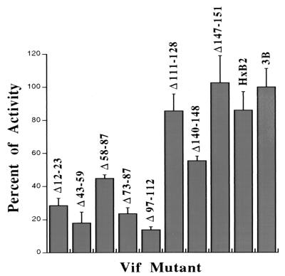
Effects of inactivating small deletion mutations in Vif on its interaction with Sp140 in the yeast two-hybrid system. LexA-BD-Vif plasmids encoding the wild-type or mutant HIV-1 strain IIIB Vif proteins or the wild-type HXB2 strain Vif were cotransfected with the Ga14-AD-Sp140 plasmid into L40 yeast cells, and colonies were selected for growth in medium lacking tryptophan, uridine, leucine, and lysine. Three individual colonies in each plate were then tested for their rates of growth in liquid culture medium lacking histidine as well as the above-mentioned omissions and supplemented with 3 mM 3-amino-1,2,4-triazole (Sigma). Growth rates were measured in the triplicate samples by adsorption at 600 nm at 24-h intervals. The results are plotted as the averages of values from two independent assays relative to the growth rate of strain IIIB, considered to be standard, and with the error bars showing the ranges of the two assays. The two-hybrid interaction enables cell replication in the absence of histidine.
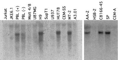
Detection of Sp140 mRNA by Northern blot analyses of RNAs from diverse human leukemic cell lines and from PBLs. RNAs were extracted from cultured cells and analyzed as previously described (50) by using a fragment of Sp140 cDNA corresponding to nucleotides 1350 to 2198 of the full-length cDNA (4) as the hybridization probe. Equal loadings of the lanes were verified by observing the quenching of autofluorescence caused by rRNAs, and sample integrity was shown by probing for the S2 ribosomal protein mRNA (results not shown). The cell lines were from the American Type Culture Collection (Manassas, Va.) or were generously provided by the AIDS Research and Reference Reagent Program at the National Institutes of Health. The P and NP phenotypes of the cell lines in the right panel were tested as previously described (30). The following pairs of cell lines had a common origin: Jurkat and JKE6.1, H9 and HUT78, and CEM-SS and A3.01. Moreover, C8166 cells were previously reported to be P or semipermissive (13, 47), implying a difference in our C8166-45 sample perhaps caused by divergence or contamination. If we consider only the other cells that were independently derived, there was a precise correspondence of the NP phenotype with the presence of Sp140 mRNA (n = 12 cell lines; P ≤ 0.001).
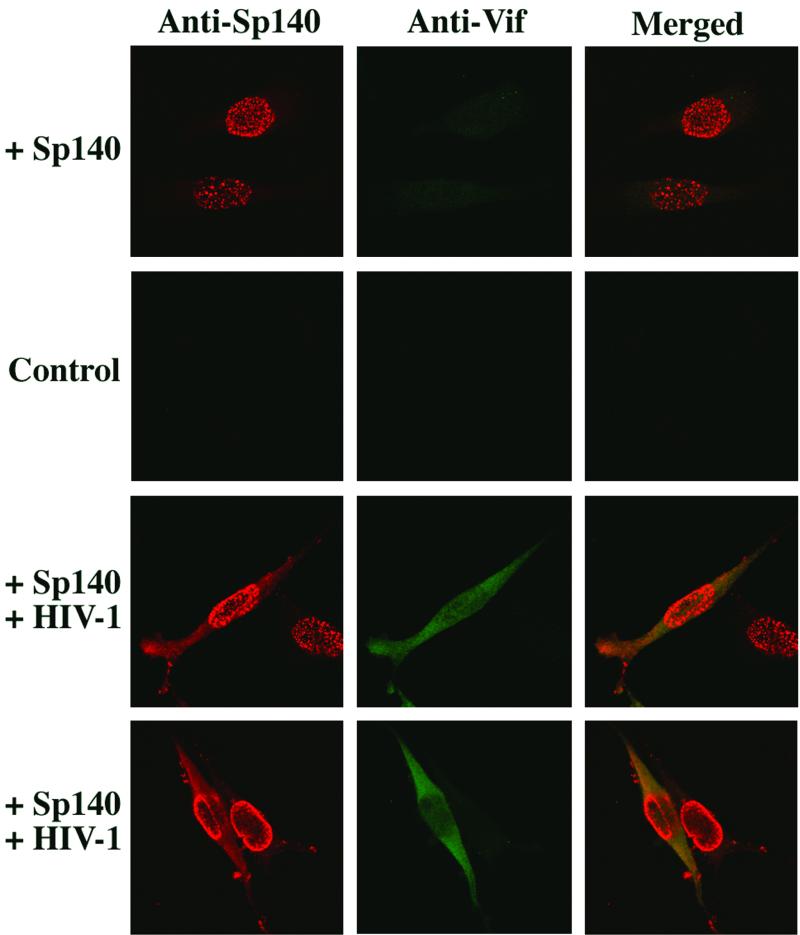
Confocal immunofluorescence microscopy of HeLa-CD4 cells expressing Sp140 in the presence or absence of Vif. The HeLa-CD4 cells (clone HTC.15) were transduced with an Ad-Sp140 expression vector (3-5) and were then infected where indicated with wild-type NL4-3 strain HIV-1 (30) for 24 h prior to fixation and staining with a rat antiserum to Sp140 (3-5). The cells were then treated with Alexa 594-conjugated anti-rat immunoglobulin (Molecular Probes, Eugene, Oreg.) or with rabbit antiserum to Vif followed by Alexa-488 conjugated secondary antibody from the same source. Confocal microscopy was done as previously described (28).
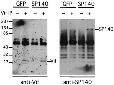
Coimmunoprecipitation of a Vif-Sp140 complex from cellular extracts. HeLa-CD4 cells were transduced with Ad-Sp140 or with a negative control Ad-GFP vector (3-5) and were then infected with wild-type HIV-1 (NL 4-3 strain) for 24 h as described in the legend for Fig. 2. The cells were then lysed with 1% Triton X-100 in 150 mM NaCl and 10 mM Tris-HCl in the presence of protease inhibitors (Sigma) at 0°C. After being centrifuged to remove nuclei and debris, the extracts were incubated with preimmune rabbit serum (1:200) and with protein A-Sepharose beads (30 μl of a 50% slurry/ml of lysate) for 1 h prior to centrifugation at 4,000 rpm for 1 min. Aliquots of the supernatants were then incubated either with more preimmune rabbit serum or with Vif antiserum prior to the addition of more protein A-Sepharose beads and recentrifugation. The pelleted beads were washed, and the extracted proteins were analyzed by electrophoresis and Western immunoblotting by using either antiserum to Vif (left panel) or antiserum to Sp140 (right panel) in comparison with protein standards as described previously (30). IP, immunoprecipitation.
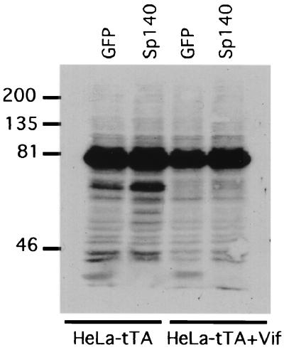
Effects of Sp140 and Vif on protein sumoylation. HeLa-tTA cells or HeLa-tTA Vif cells that express a previously described tetracycline-repressible Vif vector (30) were transduced for 24 h with equal titers of either Ad-Sp140 or Ad-GFP, which encodes GFP in the absence of tetracycline. Cell extracts were used for Western immunoblotting with a SUMO-1-specific monoclonal antibody (αGMP-1) (Zymed, Inc., South San Francisco, Calif.). Sp140 reproducibly induced elevated expression of at least one protein (Mr, ∼65,000) that reacted with the monoclonal antibody and of several minor components, and in some cases it also reduced expression of a smaller protein (Mr, ∼30,000). The inductive effects of Sp140 were eliminated by expression of Vif.
Similar articles
-
Cellular and viral specificities of human immunodeficiency virus type 1 vif protein.J Virol. 2000 Jul;74(13):5982-7. doi: 10.1128/jvi.74.13.5982-5987.2000. J Virol. 2000. PMID: 10846079 Free PMC article.
-
Human immunodeficiency virus type 1 Vif does not influence expression or virion incorporation of gag-, pol-, and env-encoded proteins.J Virol. 1996 Dec;70(12):8263-9. doi: 10.1128/JVI.70.12.8263-8269.1996. J Virol. 1996. PMID: 8970945 Free PMC article.
-
Human immunodeficiency virus type 1 Vif protein is an integral component of an mRNP complex of viral RNA and could be involved in the viral RNA folding and packaging process.J Virol. 2000 Sep;74(18):8252-61. doi: 10.1128/jvi.74.18.8252-8261.2000. J Virol. 2000. PMID: 10954522 Free PMC article.
-
The Vif protein of human immunodeficiency virus type 1 (HIV-1): enigmas and solutions.Curr Med Chem. 2004 Jan;11(2):221-31. doi: 10.2174/0929867043456124. Curr Med Chem. 2004. PMID: 14754418 Review.
-
The viral infectivity factor (Vif) of HIV-1 unveiled.Trends Mol Med. 2004 Jun;10(6):291-7. doi: 10.1016/j.molmed.2004.04.008. Trends Mol Med. 2004. PMID: 15177194 Review.
Cited by
-
Post-GWAS functional characterization of susceptibility variants for chronic lymphocytic leukemia.PLoS One. 2012;7(1):e29632. doi: 10.1371/journal.pone.0029632. Epub 2012 Jan 3. PLoS One. 2012. PMID: 22235315 Free PMC article.
-
Immune chromatin reader SP140 regulates microbiota and risk for inflammatory bowel disease.Cell Host Microbe. 2022 Oct 12;30(10):1370-1381.e5. doi: 10.1016/j.chom.2022.08.018. Epub 2022 Sep 20. Cell Host Microbe. 2022. PMID: 36130593 Free PMC article.
-
SP140L, an Evolutionarily Recent Member of the SP100 Family, Is an Autoantigen in Primary Biliary Cirrhosis.J Immunol Res. 2015;2015:526518. doi: 10.1155/2015/526518. Epub 2015 Aug 11. J Immunol Res. 2015. PMID: 26347895 Free PMC article.
-
The human immunodeficiency virus type 1 Vif protein reduces intracellular expression and inhibits packaging of APOBEC3G (CEM15), a cellular inhibitor of virus infectivity.J Virol. 2003 Nov;77(21):11398-407. doi: 10.1128/jvi.77.21.11398-11407.2003. J Virol. 2003. PMID: 14557625 Free PMC article.
-
The Speckled Protein (SP) Family: Immunity's Chromatin Readers.Trends Immunol. 2020 Jul;41(7):572-585. doi: 10.1016/j.it.2020.04.007. Epub 2020 May 5. Trends Immunol. 2020. PMID: 32386862 Free PMC article. Review.
References
-
- Bernier-Villamor, V., D. A. Sampson, M. J. Matunis, and C. D. Lima. 2002. Structural basis for E2-mediated SUMO conjugation revealed by a complex between ubiquitin-conjugating enzyme Ubc9 and RanGAP1. Cell 108:345-356. - PubMed
-
- Bloch, D. B., S. M. de la Monte, P. Guigaouri, A. Filippov, and K. D. Bloch. 1996. Identification and characterization of a leukocyte-specific component of the nuclear body. J. Biol. Chem. 271:29198-29204. - PubMed
Publication types
MeSH terms
Substances
Grants and funding
LinkOut - more resources
Full Text Sources
Other Literature Sources
Molecular Biology Databases
Miscellaneous
