. 2002 Sep 20:3:28.
doi: 10.1186/1471-2091-3-28.

MPN+, a putative catalytic motif found in a subset of MPN domain proteins from eukaryotes and prokaryotes, is critical for Rpn11 function

Affiliations

MPN+, a putative catalytic motif found in a subset of MPN domain proteins from eukaryotes and prokaryotes, is critical for Rpn11 function

Vered Maytal-Kivity et al. BMC Biochem. .

Abstract

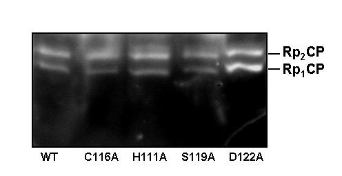
Background: Three macromolecular assemblages, the lid complex of the proteasome, the COP9-Signalosome (CSN) and the eIF3 complex, all consist of multiple proteins harboring MPN and PCI domains. Up to now, no specific function for any of these proteins has been defined, nor has the importance of these motifs been elucidated. In particular Rpn11, a lid subunit, serves as the paradigm for MPN-containing proteins as it is highly conserved and important for proteasome function.

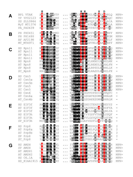
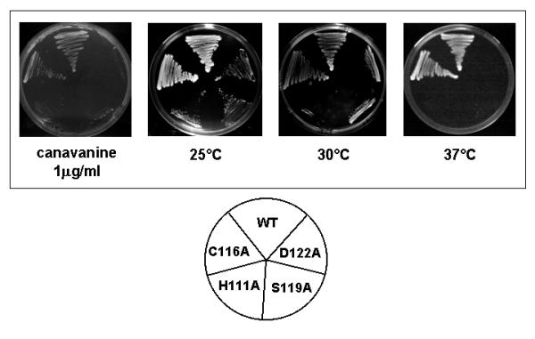
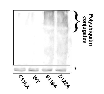
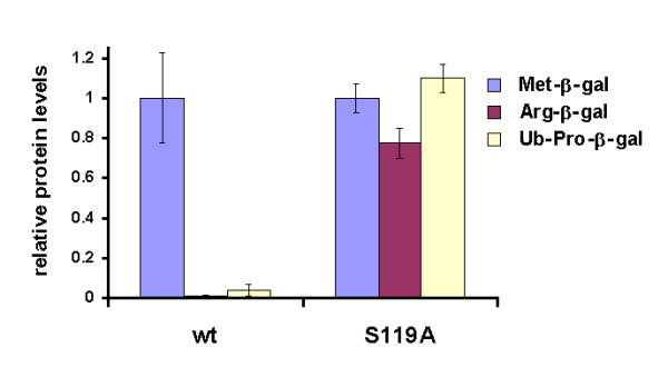
Results: We have identified a sequence motif, termed the MPN+ motif, which is highly conserved in a subset of MPN domain proteins such as Rpn11 and Csn5/Jab1, but is not present outside of this subfamily. The MPN+ motif consists of five polar residues that resemble the active site residues of hydrolytic enzyme classes, particularly that of metalloproteases. By using site-directed mutagenesis, we show that the MPN+ residues are important for the function of Rpn11, while a highly conserved Cys residue outside of the MPN+ motif is not essential. Single amino acid substitutions in MPN+ residues all show similar phenotypes, including slow growth, sensitivity to temperature and amino acid analogs, and general proteasome-dependent proteolysis defects.

Conclusions: The MPN+ motif is abundant in certain MPN-domain proteins, including newly identified proteins of eukaryotes, bacteria and archaea thought to act outside of the traditional large PCI/MPN complexes. The putative catalytic nature of the MPN+ motif makes it a good candidate for a pivotal enzymatic function, possibly a proteasome-associated deubiquitinating activity and a CSN-associated Nedd8/Rub1-removing activity.


Figures

Figure 1
Figure 1
The MPN+ motif. Sequence alignment of representative MPN domain protein, only two conserved blocks containing the MPN+ motif are shown. The proteins are grouped in seven categories: A, bacterial; B, archaeal; C, proteasome lid components; D, CSN components; E, eIF3 components; F, Prp8-like proteins; G other eukaryotic MPN proteins. In the eukaryotic groups, representative sequences from human (HS), Arabidopsis (AT), S. pombe (SP) and S. cerevisiae (SC) are shown. Prokaryotic species shown are lambda phage (BPL), Yersinia pestis (YP), Synechocystis sp. (SS), Mycobacterium tuberculosum (MyT), Pseudomonas aeruginosa (PA), Pyrococcus horikoshii (PH), Archaeoglobus fulgidus (AF), Methanobacter thermoautotrophicum (MT). Residues invariant or conservatively substituted in at least 50% of all sequences are shown on black and grey background, respectively. The MPN+ motif residues are shown in red. The top line indicates the amino acids constituting the MPN+ motif; the rightmost column indicates whether a protein is considered an MPN+ protein or a plain MPN protein.
Figure 2
Figure 2
Growth of rpn11 Mutants under Different Growth Conditions. WT and mutant strains were streaked on YPD at 25, 30 and 37°C. Cells were also plated on complete minimal media containing 1 μg/ml of the amino acid analog canavinine instead of arginine. Plates were photographed after 3–5 days. Three single amino acid substitutions in the MPN+ motif of Rpn11 were studied: his111ala, Ser119ala, and asp122ala. For comparison, a substitution of a highly conserved cysteine residue (cys116ala) that is not part of the MPN+ motif (see Fig. 1) was included as well. At 25°C, all MPN+ mutations are viable but slow growing. Hiss111 is extremely temperature sensitive, showing no appreciable growth even when shifted to 30°C, or in the presence of even 1 μg/ml of the amino acid analog canavinine. Asp122 and Ser119 substitutions show lethality under elevated temperature (37°C) or exposure to canavanine. In comparison, Cys116 substitutions show no growth defects under these conditions.
Figure 3
Figure 3
Accumulation of multiubiquitinated proteins in MPN motif mutants. Total cell extracts from WT yeast and from strains containing the MPN+ motif substitutions were separated on an 8% SDS gel and blotted with anti-ubiquitin antibodies. Cells were rapidly lysed in presence of 12% TCA in order to inhibit post lysis enzymatic activity. Accumulation of high MW polyubiquitinated proteins is detected in the Ser119 and Asp122 mutants, but not in WT or in the Cys116 mutants. His111 mutants accumulate polyubiquitinated proteins as well (not shown). A protein band migrating at around 20 kDa that is detected with the anti Ub antibody is used as an internal loading control.
Figure 4
Figure 4
Stabilization of short-lived proteasome substrates. WT and the S119A mutant strains containing the Ub-Met-, Ub-Arg-, and Ub-Pro-βgal constructs on multi-copy plasmids under the GAL promotor were tested for LacZ activity after galactose-induction. LacZ activity is indicative of steady-state levels of the reporter protein. WT cells accumulate high levels of the stable Met-βgal, but rapidly degrade Arg-βgal and UB-Pro-βgal (left panel), whereas the MPN+ mutation shows dramatic stabilization and accumulation of both short-lived fusion proteins (right panel).
Figure 5
Figure 5
Migration pattern of proteasome from WT and rpn11 mutants by non-denaturing PAGE. Cell extracts were prepared from Logarithmically growing WT or the rpn11 MPN+ mutants that were brought to an identical OD600 level. Extracts were clarified by centrifugation, and identical amounts of total protein were separated on native gel. The gel was then incubated for 10 minutes with the fluorescent peptide LLVY-AMC at 30°C, and photographed under UV light (380/440 nm). Fluorescent bands indicate migration of Doubly capped and Singly capped 26S proteasome forms [14]. There is no noticeable change in migration pattern or overall levels of proteasomes from the different rpn11 mutants, indicating that mutated Rpn11 is properly incorporated into the lid and no gross structural changes in proteasome composition or amounts are due to the mutations.

Similar articles

Cited by

References

    1. Pickart CM. Mechanisms underlying ubiquitination. Annu Rev Biochem. 2001;70:503–533. doi: 10.1146/annurev.biochem.70.1.503. - DOI - PubMed
    1. Glickman MH, Ciechanover A. The Ubiquitin-proteasome Proteolytic Pathway: Destruction for the sake of construction. Physiol Rev. 2002;82:373–428. - PubMed
    1. Chung CH, Baek SH. Deubiquitinating enzymes: Their diversity and emerging roles. Biochem Biophys Res Commun. 1999;266:633–640. doi: 10.1006/bbrc.1999.1880. - DOI - PubMed
    1. D'Andrea A, Pellman D. Deubiquitinating enzymes: A new class of biological regulators. Crit Rev Biochem Mol Biol. 1998;33:337–332. - PubMed
    1. Glickman MH, Rubin DM, Coux O, Wefes I, Pfeifer G, Cjeka Z, Baumeister W, Fried VA, Finley D. A subcomplex of the proteasome regulatory particle required for ubiquitin-conjugate degradation and related to the COP9/Signalosome and eIF3. Cell. 1998;94:615–623. - PubMed

Publication types

MeSH terms