Review
. 2002 Oct;3(10):944-50.
doi: 10.1093/embo-reports/kvf202.

Structural organization of the endoplasmic reticulum

Affiliations
Review

Structural organization of the endoplasmic reticulum

Gia K Voeltz et al. EMBO Rep. 2002 Oct.

Abstract

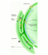
The endoplasmic reticulum (ER) is a continuous membrane system but consists of various domains that perform different functions. Structurally distinct domains of this organelle include the nuclear envelope (NE), the rough and smooth ER, and the regions that contact other organelles. The establishment of these domains and the targeting of proteins to them are understood to varying degrees. Despite its complexity, the ER is a dynamic structure. In mitosis it must be divided between daughter cells and domains must be re-established, and even in interphase it is constantly rearranged as tubules extend along the cytoskeleton. Throughout these rearrangements the ER maintains its basic structure. How this is accomplished remains mysterious, but some insight has been gained from in vitro systems.


Figures

Figure 1
Figure 1
Ultrastructure of the RER, SER and NE. (A) Localization of a GFP–ER fusion protein (GFP-Sec61β) expressed in a COS cell and visualized by epifluorescence microscopy. The fine ER network is particularly clear in the thin edges of the cell periphery; three-way junctions and polygonal reticulum are easily visualized. (B) An electron micrograph of a liver cell shows RER (rough reticulum) and patches of SER (smooth reticulum) (picture taken from Fawcett, 1966, with copyright permission from the publisher, W.B. Saunders Co.). (C) Localization of GFP–ER fusion protein (GFPsec63) expressed in yeast and visualized by fluorescence microscopy outlines the structure of the ER (the upper picture focuses on the middle of the cell, the lower on the top of the cell; taken from Prinz et al., 2001, with copyright permission of the Rockefeller University Press). Several tubules (arrow in top panel) connect the outer NE (top panel) to the peripheral ER (bottom panel).
Figure 2
Figure 2
Targeting and retention of an inner nuclear membrane (INM) protein. (A) The newly translated protein is translocated into the ER membrane. (B) It diffuses through the peripheral ER. (C) It then diffuses through the nuclear pore membrane to the inner nuclear envelope. (D) Retention of the INM protein as a result of its binding to the nuclear lamina (green) and chromatin (yellow).
None
None
None

References

    1. Allan V.J. and Vale R.D. (1991) Cell cycle control of microtubule-based membrane transport and tubule formation in vitro. J. Cell Biol., 113, 346–359. - PMC - PubMed
    1. Amar-Cortesec A., Dublet B. and Beaufay H. (1989) Translocation and proteolytic processing of nascent secretory polypeptide chains: two functions associated with the ribosomal domain of the endoplasmic reticulum. Mol. Biol. Cell, 65, 99–108. - PubMed
    1. Bailer S.M., Eppenberger H.M., Griffiths G. and Nigg E.A. (1991) Characterization of a 54-kD protein of the inner nuclear membrane: evidence for cell cycle-dependent interaction with the nuclear lamina. J. Cell Biol., 114, 389–400. - PMC - PubMed
    1. Banta M., Polizotto R.S., Wood S.A., de Figueiredo P. and Brown W.J. (1995) Characterization of a cytosolic activity that induces the formation of Golgi membrane tubules in a cell-free reconstitution system. Biochemistry, 34, 13359–13366. - PubMed
    1. Baumann O. and Walz B. (2001) Endoplasmic reticulum of animal cells and its organization into structural and functional domains. Int. Rev. Cytol., 205, 149–214. - PubMed

Publication types

Substances

LinkOut - more resources