. 2002 Oct 14;159(1):113-22.
doi: 10.1083/jcb.200202075. Epub 2002 Oct 7.

The LIM-only protein FHL2 interacts with beta-catenin and promotes differentiation of mouse myoblasts

Affiliations

The LIM-only protein FHL2 interacts with beta-catenin and promotes differentiation of mouse myoblasts

Bernd Martin et al. J Cell Biol. .

Abstract

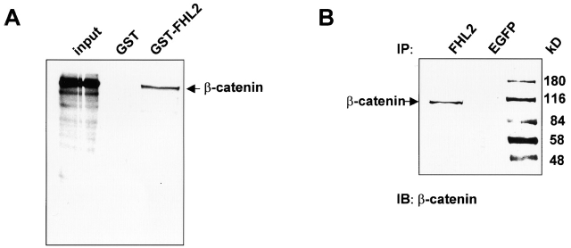
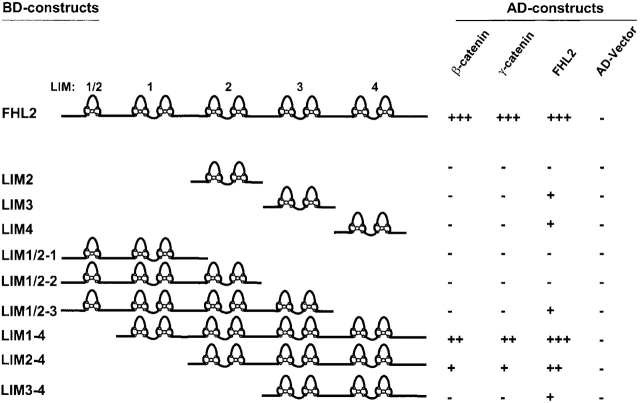
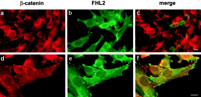
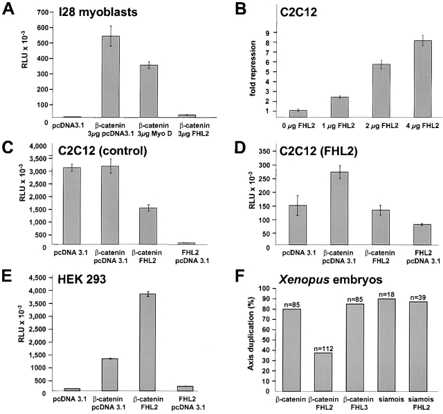
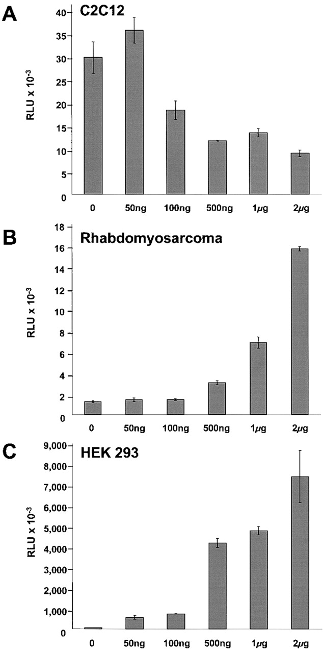
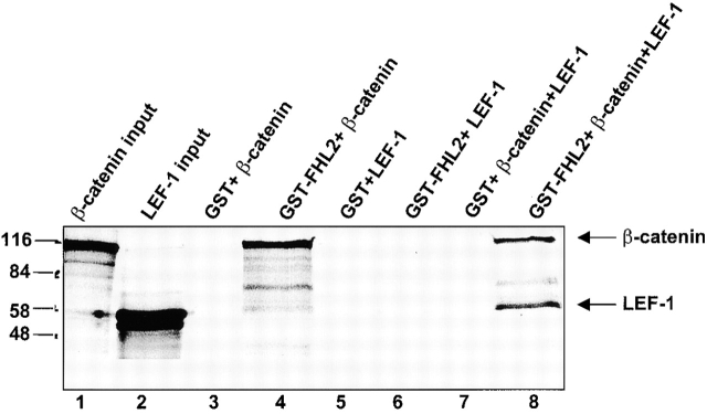
FHL2 is a LIM-domain protein expressed in myoblasts but down-regulated in malignant rhabdomyosarcoma cells, suggesting an important role of FHL2 in muscle development. To investigate the importance of FHL2 during myoblast differentiation, we performed a yeast two-hybrid screen using a cDNA library derived from myoblasts induced for differentiation. We identified beta-catenin as a novel interaction partner of FHL2 and confirmed the specificity of association by direct in vitro binding tests and coimmunoprecipitation assays from cell lysates. Deletion analysis of both proteins revealed that the NH2-terminal part of beta-catenin is sufficient for binding in yeast, but addition of the first armadillo repeat is necessary for binding FHL2 in mammalian cells, whereas the presence of all four LIM domains of FHL2 is needed for the interaction. Expression of FHL2 counteracts beta-catenin-mediated activation of a TCF/LEF-dependent reporter gene in a dose-dependent and muscle cell-specific manner. After injection into Xenopus embryos, FHL2 inhibited the beta-catenin-induced axis duplication. C2C12 mouse myoblasts stably expressing FHL2 show increased myogenic differentiation reflected by accelerated myotube formation and expression of muscle-specific proteins. These data imply that FHL2 is a muscle-specific repressor of LEF/TCF target genes and promotes myogenic differentiation by interacting with beta-catenin.


Figures

Figure 1.
Figure 1.
Interaction between FHL2 and β-catenin in vitro and in vivo. (A) Recombinant GST or GST–FHL2 fusion protein was incubated with [35S]methionine-labeled β-catenin. After extensive washing, proteins retained by GST or GST–FHL2 were analyzed together with 10% of the input material by SDS–PAGE and fluorography. (B) C2C12 cell lysates were immunoprecipitated (IP) with rabbit pAbs against FHL2 or EGFP (negative control) as indicated and analyzed by SDS-PAGE and immunoblotting (IB) using the anti–β-catenin mAb 14. The arrows show coprecipitated β-catenin.
Figure 2.
Figure 2.
Specificity of interaction of FHL proteins with α-catenin, β-catenin, and γ-catenin. (A) Yeast Y190 cells were cotransformed with GAL4-DNA–binding domain (BD) and GAL4–activation domain (AD) chimeric constructs. The interaction was evaluated using a β-Gal filter assay as described in Materials and methods. The interactions of FHL proteins with AD-FHL2 or the empty AD-vector represent positive or negative controls. (B) HEK 293 cells were transiently transfected with cDNA constructs as indicated. After 34 h, RIPA cell lysates were divided into two parts and proteins were either precipitated (P) with glutathione-conjugated Sepharose beads for GST-tagged proteins or immunoprecipitated (IP) with the antibody 9E10 for myc-tagged proteins. The coprecipitated proteins were analyzed in immunoblots (IB) with appropriate antibodies. After the first immunodetection, the blots were stripped and redeveloped with antibodies against GST- or myc-tag to ascertain the amount of precipitated proteins.
Figure 3.
Figure 3.
The NH 2 terminus of β-catenin is sufficient for binding to FHL2. (A) Yeast Y190 cells were cotransformed with GAL4-DNA–binding domain (FHL2) and GAL4–activation domain (β-catenin) chimeric constructs, and protein–protein interactions were evaluated by a β-Gal filter assay. The numbers over the diagrams indicate β-catenin amino acids encoded by each construct. The arrangement of armadillo repeats is based on Hulsken et al. (1994). (B) HEK 293 cells were transiently transfected with cDNA constructs as indicated and lysed with RIPA buffer. The presence of myc-tagged β-catenin and its deletion mutants in GST–FHL2 precipitates (P) were detected by immunoblotting (IB) with myc antibody (top blot). To ascertain the amount of precipitated FHL2 protein, the blot was redeveloped with antibodies against GST. The blot on the bottom shows the expression in transfected cells of full-length myc β-catenin and deletion mutants; β-catenin (NT-1) with the NH2 terminus plus the first armadillo repeat, and β-catenin (NT) with the NH2 terminus only. 10 μg of total cell lysates were loaded.
Figure 3.
Figure 3.
The NH 2 terminus of β-catenin is sufficient for binding to FHL2. (A) Yeast Y190 cells were cotransformed with GAL4-DNA–binding domain (FHL2) and GAL4–activation domain (β-catenin) chimeric constructs, and protein–protein interactions were evaluated by a β-Gal filter assay. The numbers over the diagrams indicate β-catenin amino acids encoded by each construct. The arrangement of armadillo repeats is based on Hulsken et al. (1994). (B) HEK 293 cells were transiently transfected with cDNA constructs as indicated and lysed with RIPA buffer. The presence of myc-tagged β-catenin and its deletion mutants in GST–FHL2 precipitates (P) were detected by immunoblotting (IB) with myc antibody (top blot). To ascertain the amount of precipitated FHL2 protein, the blot was redeveloped with antibodies against GST. The blot on the bottom shows the expression in transfected cells of full-length myc β-catenin and deletion mutants; β-catenin (NT-1) with the NH2 terminus plus the first armadillo repeat, and β-catenin (NT) with the NH2 terminus only. 10 μg of total cell lysates were loaded.
Figure 4.
Figure 4.
Identification of the minimal binding site on FHL2 for β-catenin and γ-catenin by yeast two-hybrid assays. Yeast Y190 cells were cotransformed with GAL4-DNA–binding domain and GAL4–activation domain chimeric constructs and protein–protein interactions were evaluated by a β-Gal filter assay. The known interaction of FHL2 with its own subdomains was used as a positive control, and the empty AD-vector served as a negative control.
Figure 5.
Figure 5.
Subcellular localization of FHL2 and β-catenin. C2C12 cells cultured on coverslips were transiently transfected with EGFP–FHL2. Cells immunostained with anti-β–catenin mAb and Alexa-conjugated goat anti–mouse polyclonal antibody were analyzed by fluorescence microscopy. Although β-catenin (red) is concentrated at the membrane and at cell–cell contacts, EGFP–FHL2 (green) is concentrated at focal adhesion sites. The merged images show no colocalization of the two proteins at membrane level. Panels a–c and d– f represent pictures taken with 40× or 63× objectives, respectively. Bars, ∼10 μm.
Figure 6.
Figure 6.
FHL2 influences β-catenin–mediated activation of TCF/LEF-dependent transcription. (A) TOPflash luciferase reporter plasmid was cotransfected with β-catenin S33A and the indicated amounts of FHL2 or MyoD (as a control) cDNAs into I28 mouse myoblast cells. Luciferase activity is given in relative light units (RLU). Basal activity of the TOPflash promoter was measured in the presence of empty expression vector to maintain equivalent amounts of transfected DNA. Results shown are the average of three independent transfections. (B) C2C12 cells were cotransfected with constant amounts of TOPflash plasmid and β-catenin S33A, and with increasing amounts of FHL2 plasmid as indicated. Values indicate fold repression of the β-catenin–induced reporter gene activity. The induction of reporter gene activity by β-catenin alone was arbitrarily taken as unity. (C–E) C2C12 cells stably infected with pBabe vector alone (C), or pBabe myc-FHL2 vector (D), or HEK 293 cells (E) were transfected with TOPflash luciferase reporter plasmid and the indicated expression plasmids. (F) Xenopus embryos were injected with different RNAs as indicated at the four-cell stage. Bars represent the percentage of embryos with duplicated axis at stage 19–23. n, No. of embryos examined.
Figure 7.
Figure 7.
FHL2 exhibits an autonomous repression function restricted to fusion-competent myoblasts. Increasing amounts of GAL-FHL2 plasmid and 1μg 5xUAS tk-luc reporter plasmid were cotransfected as indicated into C2C12 (A), Rhabdomyosarcoma (B), or HEK 293 cells (C).
Figure 8.
Figure 8.
β-Catenin forms a ternary protein complex with FHL2 and LEF-1. Recombinant GST–FHL2 (lanes 4, 6, and 8) or GST (lanes 3, 5, and 7) proteins were incubated with [35S]methionine-labeled β-catenin (lanes 3 and 4) or [35S]methionine-labeled LEF-1 (lanes 5 and 6), or with both (lanes 7 and 8). After extensive washing, proteins retained by GST or GST–FHL2 were analyzed together with 10% of the input material (lanes 1 and 2) by SDS–PAGE and fluorography.
Figure 9.
Figure 9.
Ectopic expression of FHL2 promotes differentiation of C2C12 myoblasts. (A) C2C12 myoblasts infected with retroviruses containing the pBabe vector alone (control) or pBabe with myc-FHL2 (FHL2) were cultured in growth medium until 90–95% confluency (day 0) and then shifted to differentiation medium (days 1–5). (B) Lysates (20 μg protein/lane) from cells cultured in growth medium (day 0) or in differentiation medium (days 1–5) were immunoblotted with anti-myc mAb 9E10, anti-FHL2 mAb, anti–β-catenin mAb 14, anti-MyHC mAb NOQ7.5.4D, or anti–cyclin D1 mAb DCS-6.

References

    1. Aberle, H., A. Bauer, J. Stappert, A. Kispert, and R. Kemler. 1997. β-Catenin is a target for the ubiquitin-proteasome pathway. EMBO J. 16:3797–3804. - PMC - PubMed
    1. Akiyama, T. 2000. Wnt/β-catenin signaling. Cytokine Growth Factor Rev. 11:273–282. - PubMed
    1. Baniahmad, A., X. Leng, T.P. Burris, S.Y. Tsai, M.J. Tsai, and B.W. O'Malley. 1995. The τ4 activation domain of the thyroid hormone receptor is required for release of a putative corepressor(s) necessary for transcriptional silencing. Mol. Cell. Biol. 15:76–86. - PMC - PubMed
    1. Beckerle, M.C. 1997. Zyxin: zinc fingers at sites of cell adhesion. Bioessays. 19:949–957. - PubMed
    1. Behrens, J., J.P. von Kries, M. Kuhl, L. Bruhn, D. Wedlich, R. Grosschedl, and W. Birchmeier. 1996. Functional interaction of β-catenin with the transcription factor LEF-1. Nature. 382:638–642. - PubMed

Publication types

MeSH terms