. 2002 Oct 7;196(7):923-34.
doi: 10.1084/jem.20020774.

Regulation of the subcellular localization of tumor necrosis factor receptor-associated factor (TRAF)2 by TRAF1 reveals mechanisms of TRAF2 signaling

Affiliations

Regulation of the subcellular localization of tumor necrosis factor receptor-associated factor (TRAF)2 by TRAF1 reveals mechanisms of TRAF2 signaling

Joseph R Arron et al. J Exp Med. .

Abstract

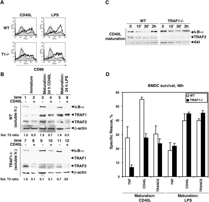
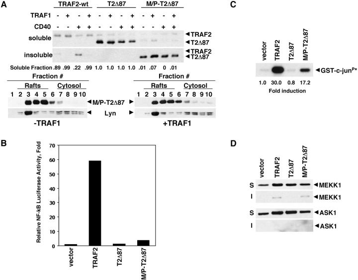
Tumor necrosis factor receptor-associated factor (TRAF)2 is a critical adaptor molecule for tumor necrosis factor (TNF) receptors in inflammatory and immune signaling. Upon receptor engagement, TRAF2 is recruited to CD40 and translocates to lipid rafts in a RING finger-dependent process, which enables the activation of downstream signaling cascades including c-Jun NH(2)-terminal kinase (JNK) and nuclear factor (NF)-kappaB. Although TRAF1 can displace TRAF2 and CD40 from raft fractions, it promotes the ability of TRAF2 activate signaling over a sustained period of time. Removal of the RING finger of TRAF2 prevents its translocation into detergent-insoluble complexes and renders it dominant negative for signaling. TRAF1(-/-) dendritic cells show attenuated responses to secondary stimulation by TRAF2-dependent factors and increased stimulus-dependent TRAF2 degradation. Replacement of the RING finger of TRAF2 with a raft-targeting signal restores JNK activation and association with the cyto-skeletal protein Filamin, but not NF-kappaB activation. These findings offer insights into the mechanism of TRAF2 signaling and identify a physiological role for TRAF1 as a regulator of the subcellular localization of TRAF2.


Figures

Figure 1.
Figure 1.
TRAF1 regulates the detergent solubility of TRAF2. (A) 293T HEK cells were transfected in 6-well plates with the indicated amounts of CD40, TRAF1, and TRAF2. Total DNA content was maintained constant at 1 μg by the addition of empty vector. Cells were lysed in 0.75% Triton X-100, and soluble (S) and insoluble (I) fractions were immunoblotted as indicated. After probing with TRAF2 antibodies (C-20), blots were stripped and reprobed with anti-Flag M2 to detect TRAF1 and CD40. (B) As in panel A, but without transfection of CD40. hTNF-α (10 ng/ml) was added to the culture medium 6 h before harvesting. (C) As in panel A. TRAF2 was transfected in the amounts indicated. (D) As in panel A, but with 0.1 μg of TRAF2 or an NH2-terminal truncation mutant removing the first 87 residues (comprising the RING finger) of TRAF2 (T2Δ87). 0.5 μg of TRAF1 was transfected where indicated (+). (E) As in panel A. (F) 293T cells were transfected with 1.5 μg of TRAF2 or T2Δ87, 2.5 μg of TRAF1, and 1.0 μg of CD40 where indicated. Cells were treated with CD40L 6 h before harvesting then subjected to sucrose gradient density centrifugation as described in Materials and Methods and immunoblotted as indicated.
Figure 2.
Figure 2.
TRAF2 regulates the steady-state detergent solubility of CD40. (A) 293T cells were transfected with Flag-tagged CD40 constructs (0.5 μg of WT or mutants as indicated) and TRAF2 (0.5 μg, top) or Flag-tagged TRAF6 (0.5 μg, bottom). CD40 was immunoprecipitated with anti-Flag M2 antibodies and immunoprecipitates were probed for TRAF2 and CD40 as indicated. TRAF6 was immunoprecipitated with anti-TRAF6 antibodies and immunoprecipitates were probed for CD40 and TRAF6 as indicated. (B) As in Fig. 1 F, but cells were transfected with CD40-WT or the indicated mutants (1.0 μg) and TRAF2 or TRAF6 (1.5 μg) and subjected to sucrose density gradient centrifugation and immunoblotting.
Figure 3.
Figure 3.
TRAF1 promotes sustained TRAF2-mediated JNK and NF-κB activation. (A) 293T cells were transfected with TRAF2 (100 ng) and TRAF 1 (0, 33, 100, 300, or 900 ng, indicated by broadening line) as indicated. 6 h before harvesting, 10 ng/ml TNF-α was added to the culture medium. Cells were lysed in 0.75% Triton X-100 and subjected to an in vitro JNK kinase assay. (B) 293T cells were transfected with varying amounts of TRAF1 (0, 100, or 500 ng, indicated by broadening line), CD40 (50 ng), and TRAF2 (100 ng) as indicated and subjected to an NF-κB reporter assay. Values are indicated as fold increase over background, and are normalized against an internal standard (β-galactosidase).
Figure 4.
Figure 4.
TRAF1−/− DCs have deficient secondary responses to TRAF2-dependent signals. (A) Wild-type and TRAF1−/− (T1−/−) DCs were matured by overnight culture in CD40L (1:200) or LPS (100 ng/ml) and CD86 expression was monitored by FACS® analysis. Immature cells are shown as shaded areas on the histogram and matured cells are shown as broad dark lines. (B) DCs were matured as in panel A (immature, lanes 1–2 and 7–8; CD40L matured, lanes 3–4 and 9–10; LPS matured, lanes 5–6 and 11–12), starved in medium containing 0.5% serum for 2 h, and restimulated with CD40L (1:200) as indicated for 20 min (even numbered lanes; odd numbered lanes were not restimulated). Cells were lysed and the soluble fractions were immunoblotted as indicated. Ratios of soluble TRAF2 relative to the level of soluble TRAF2 in unstimulated immature cells were determined by densitometry and normalized to β-actin levels, and are indicated below the β-actin blots. (C) DCs were matured in CD40L and starved as in B, then restimulated with CD40L as indicated. Cells were lysed in 1% SDS to solubilize total cellular protein and immunoblotted as indicated. Akt levels are shown as a loading control. (D) DCs prepared as in panel A were incubated in normal medium, or medium containing TNF-α (10 ng/ml), CD40L (1:200), or TRANCE (1 μg/ml) for 48 h as indicated. Survival was determined by PI exclusion FACS®. Specific rescue is represented as (% of surviving cells [stimulated] − % of surviving cells [unstimulated])/([100 − % of surviving cells [unstimulated]).
Figure 5.
Figure 5.
Forced raft localization of TRAF2 is sufficient to activate JNK, but not NF-κB. (A) Top, 0.1 μg of wild-type TRAF2 (wt), T2Δ87, or T2Δ87 with an NH2-terminal myristoylation-palmitoylation signal peptide (M/P-T2Δ87) was cotransfected with or without TRAF1 (+, 0.5 μg), with or without CD40 (+, 0.2 μg) as indicated. Soluble and insoluble fractions were prepared as in Fig. 1. The relative proportion of soluble vs. insoluble TRAF2 or its mutants in a given lane was determined by densitometry (pixel density [PD]sol/PDsol+PDinsol) and is indicated below the immunoblots (note: this is a reflection of values relative to one another, but does not provide an absolute measure of solubility). (Bottom) Cells were transfected with M/P-T2Δ87, CD40, and +/− TRAF1 and subjected to sucrose density gradient centrifugation as in Fig. 1 F. (B) Cells were transfected with the indicated TRAF2 constructs (0.4 μg) and subjected to an NF-κB reporter assay as in Fig. 3 B. (C) Cells were transfected as in B and subjected to an in vitro JNK assay as in Fig. 3 A. (D) Cells were transfected with the indicated TRAF2 constructs (0.3 μg) and MEKK1 or ASK1 (0.3 μg) as indicated and soluble and insoluble fractions were prepared and immunoblotted.
Figure 6.
Figure 6.
Interactions with TRAF2 and the actin-binding protein Filamin are dependent on raft translocation of TRAF2. Cells were transfected as indicated with HA-tagged Filamin (amino acids 1644–2118; 0.3 μg), TRAF2 constructs (0.3 μg), and TRAF1 (0.3 μg) as indicated. Upon harvesting, cells were lysed in 0.5% NP-40, subjected to immunoprecipitation of Filamin with antibodies against HA, and immunoblotted as indicated.
Figure 7.
Figure 7.
Proposed model of the mechanism of TRAF2 signaling and its regulation by TRAF1. Upon ligand engagement, a TNFR family protein recruits TRAF2 and various kinases via the COOH-terminal TRAF domain of TRAF2. The transmembrane receptor complex assembles in the detergent-soluble fraction. Upon complex assembly, the NH2-terminal RING finger of TRAF2 mediates translocation of the receptor complex into detergent-resistant lipid rafts. This translocation event simultaneously activates and releases the kinases, while isolating TRAF2 in an insoluble complex that may be internalized and/or degraded. The activated kinases ultimately activate transcription factors such as NF-κB and AP-1, which up-regulate the expression of TRAF1. TRAF1 then releases TRAF2 from insoluble complexes by hetero-oligomerization with TRAF2 or competing for receptor binding sites. This results in an increase of soluble TRAF2 that is available for subsequent signaling events mediated by other TRAF2-dependent TNFR family proteins.

References

    1. Locksley, R.M., N. Killeen, and M.J. Lenardo. 2001. The TNF and TNF receptor superfamilies: integrating mammalian biology. Cell. 104:487–501. - PubMed
    1. Wajant, H., F. Henkler, and P. Scheurich. 2001. The TNF-receptor-associated factor family: scaffold molecules for cytokine receptors, kinases and their regulators. Cell. Signal. 13:389–400. - PubMed
    1. Rothe, M., S.C. Wong, W.J. Henzel, and D.V. Goeddel. 1994. A novel family of putative signal transducers associated with the cytoplasmic domain of the 75 kDa tumor necrosis factor receptor. Cell. 78:681–692. - PubMed
    1. Park, Y.C., V. Burkitt, A.R. Villa, L. Tong, and H. Wu. 1999. Structural basis for self-association and receptor recognition of human TRAF2. Nature. 398:533–538. - PubMed
    1. Ye, H., Y.C. Park, M. Kreishman, E. Kieff, and H. Wu. 1999. The structural basis for the recognition of diverse receptor sequences by TRAF2. Mol. Cell. 4:321–330. - PubMed

Publication types

MeSH terms