. 2002 Oct 7;196(7):935-46.
doi: 10.1084/jem.20020772.

Interleukin 15 controls both proliferation and survival of a subset of memory-phenotype CD8(+) T cells

Affiliations

Interleukin 15 controls both proliferation and survival of a subset of memory-phenotype CD8(+) T cells

Adam D Judge et al. J Exp Med. .

Abstract

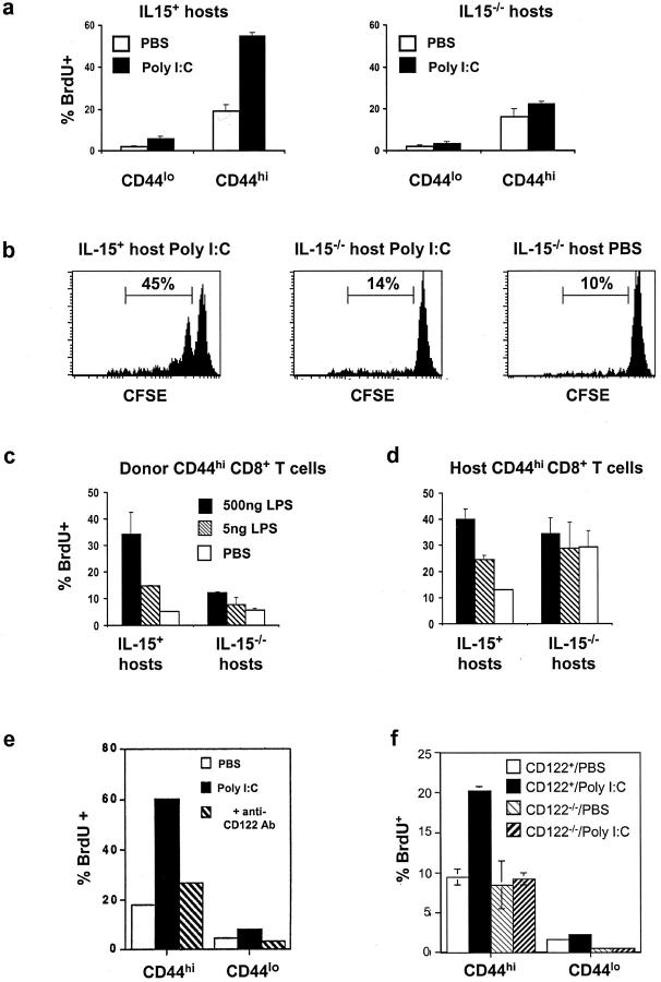
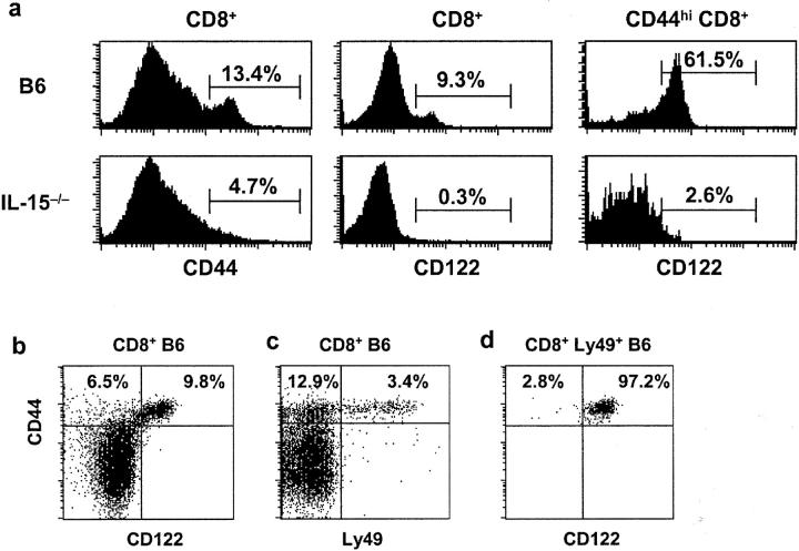
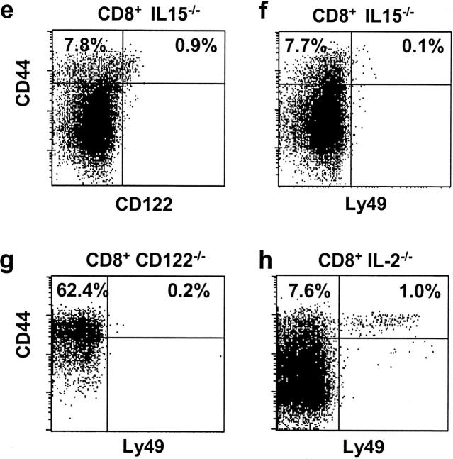
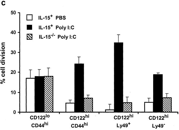
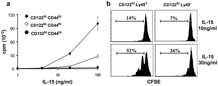
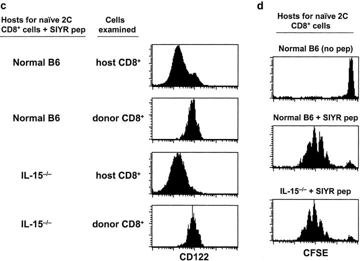
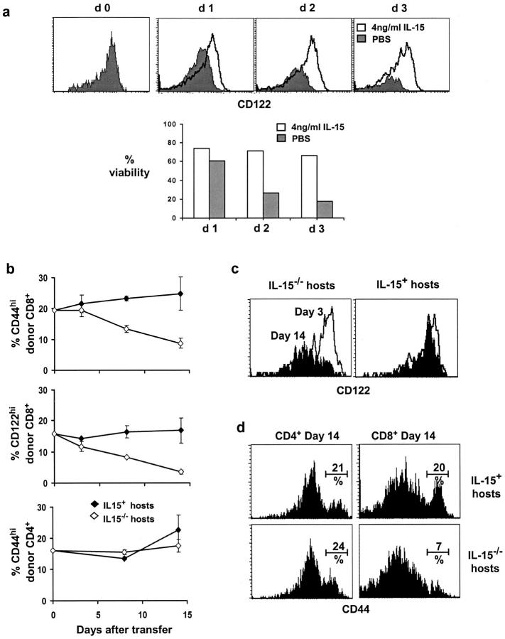
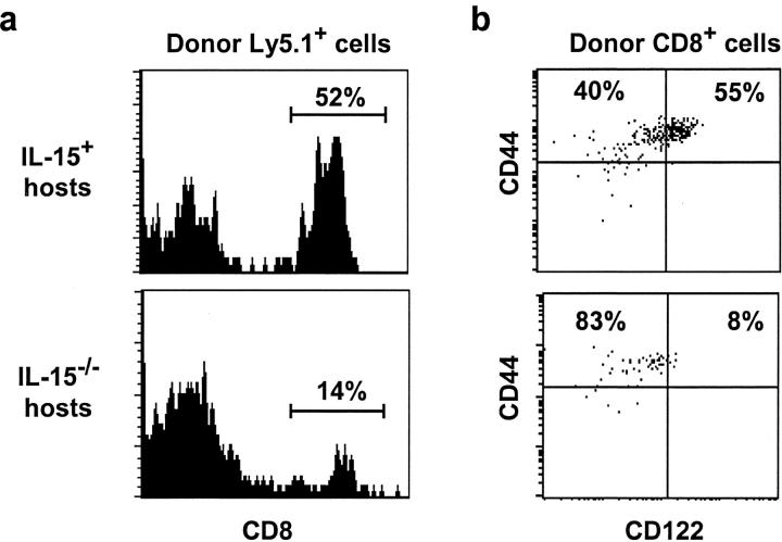
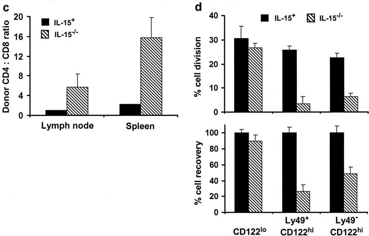
Previous work has shown that memory-phenotype CD44(hi) CD8(+) cells are controlled by a cytokine, interleukin (IL)-15. However, the dependency of CD44(hi) CD8(+) cells on IL-15 is partial rather than complete. Here, evidence is presented that CD44(hi) CD8(+) cells comprise a mixed population of IL-15-dependent and IL-15-independent cells. The major subset of CD122(hi) CD44(hi) CD8(+) cells is heavily dependent on IL-15 by three different parameters, namely (1) "bystander" proliferation induced via IFN-induced stimulation of the innate immune system, (2) normal "background" proliferation, and (3) T cell survival; IL-15 dependency is most extreme for the Ly49(+) subset of CD122(hi) CD44(hi) CD8(+) cells. In contrast to CD122(hi) cells, the CD122(lo) subset of CD44(hi) CD8(+) cells is IL-15 independent; likewise, being CD122(lo), CD44(hi) CD4(+) cells are IL-15 independent. Thus, subsets of memory-phenotype T cells differ radically in their sensitivity to IL-15.


Figures

Figure 1.
Figure 1.
Bystander proliferation is IL-15 dependent. (a and b) Purified LN T cells from Ly5.1 B6 mice were transferred to normal B6 (IL-15+) mice and IL-15−/− littermates (both Ly5.2). Hosts were then given 50 μg poly I:C intraperitoneally and proliferation was measured on day 3 by (a) BrdU incorporation after administration of BrdU given in the drinking water from day 0 or (b) dilution of CFSE by CFSE-labeled T cells; control mice received injections of saline (PBS). The data show proliferation measured on CD44hi (a and b) and CD44lo (a) subsets of gated donor (Ly5.1) CD8+ cells recovered from host LN. For CFSE labeling (b), values refer to the percentage of cells that had divided one or more times. Mean of data (± SD, four mice per group) (a) or representative data (b) are shown. Similar findings applied to cells recovered from host spleen (not shown). (c and d), as in (a) except that mice were injected with LPS, given in two different doses. The data show mean (± SD) BrdU incorporation for four mice per group for (c) donor (Ly5.1+) CD44hi CD8+ cells and (d) host (Ly5.1) CD44hi CD8+ cells. (e) Capacity of anti-CD122 mAb to inhibit proliferation induced by poly I:C. Normal B6 mice were injected with PBS, poly I:C, or a mixture of poly I:C and anti-CD122 mAb and placed on BrdU water for 3 d. The data show BrdU incorporation by gated CD44hi and CD44lo CD8+ subsets from LN pooled from three mice per group; similar results were found in another experiment. (f) Poly I:C–induced proliferation of T cells in CD122−/− → CD122+ chimeras. Irradiated (600 cGy) Thy 1.1 B6 mice were injected with T cell–depleted BM cells from CD122−/− mice on a B6 (Thy 1.2) background. After 5 mo, the chimeras were injected with poly I:C, placed on BrdU water for 3 d and stained. The data show BrdU incorporation (± SD) by gated donor CD122−/− (Thy 1.2) versus host CD122+ (Thy 1.1) CD44hi and CD44lo CD8+ cells from LN from three mice per group. Similar results were found in another experiment.
Figure 2.
Figure 2.
A subset of CD44hi CD8+ T cells is IL-15 dependent. (a) Histograms show expression of CD44 on gated CD8+ cells and CD122 on gated CD8+ and CD44hi CD8+ cells from LN of wild-type B6 versus IL-15−/− mice. (b–h) The data show CD122 or Ly49 versus CD44 expression on gated CD8+ LN T cells from wild-type B6 mice (b–d), IL-15−/− mice (e and f), CD122−/− mice (g), and IL-2−/− mice (h). The data are representative of >4 mice (aged 2–3 mo)/group and also applied to CD8+ spleen and peripheral blood cells (not shown). In d, data refer to gated Ly49+ CD8+ T cells. In all groups, Ly49 was detected using a panel of anti-Ly49A, C/I, F, and G2 mAbs that have previously been shown to be the most abundantly expressed Ly49 family members on CD8+ T cells in B6 mice (reference 20).
Figure 2.
Figure 2.
A subset of CD44hi CD8+ T cells is IL-15 dependent. (a) Histograms show expression of CD44 on gated CD8+ cells and CD122 on gated CD8+ and CD44hi CD8+ cells from LN of wild-type B6 versus IL-15−/− mice. (b–h) The data show CD122 or Ly49 versus CD44 expression on gated CD8+ LN T cells from wild-type B6 mice (b–d), IL-15−/− mice (e and f), CD122−/− mice (g), and IL-2−/− mice (h). The data are representative of >4 mice (aged 2–3 mo)/group and also applied to CD8+ spleen and peripheral blood cells (not shown). In d, data refer to gated Ly49+ CD8+ T cells. In all groups, Ly49 was detected using a panel of anti-Ly49A, C/I, F, and G2 mAbs that have previously been shown to be the most abundantly expressed Ly49 family members on CD8+ T cells in B6 mice (reference 20).
Figure 3.
Figure 3.
Bystander proliferation of CD44hi CD8+ cells is largely restricted to CD122hi subsets and most prominent for Ly49+ CD122hi cells. (a) Bystander proliferation was measured in vivo by injecting mice with graded doses of LPS, placing mice on BrdU water for 3 d followed by FACS® analysis of LN and spleen cells. Injections of PBS were used as a control. The data show BrdU incorporation by CD122hi CD44hi CD8+ cells versus CD122lo CD44hi CD8+ cells from LN; mean values (± SD) of three mice per group. In further experiments, proliferation was measured by transferring CFSE-labeled Ly5.1 B6 T cells into (b) syngenic B6 and (c) B6 versus IL-15−/− mice (both Ly5.2), injecting the hosts with 50 μg poly I:C, then using FACS® analysis to study division (loss of CFSE) by the donor cells 3 d later. In b, the data show CFSE labeling of donor-derived (Ly5.1+) CD122hi CD44hi CD8+ versus CD122lo CD44hi CD8+ subsets recovered from spleen. Cells from LN showed a similar response. The data are representative of four mice tested individually. In c, the data show the percentage of donor T cell subsets that underwent one or more rounds of division (the percentage of cell division) after poly I:C injection. Data are shown for CD44hi CD8+ spleen cells gated on CD122lo, total CD122hi, CD122hi Ly49+, and CD122hi Ly49 subsets after transfer to IL-15+ (black bars) or IL-15−/− mice (striped bars); cells transferred to PBS-injected IL-15+ mice (white bars) were used as controls. The data show mean values (± SD) for four mice per group.
Figure 3.
Figure 3.
Bystander proliferation of CD44hi CD8+ cells is largely restricted to CD122hi subsets and most prominent for Ly49+ CD122hi cells. (a) Bystander proliferation was measured in vivo by injecting mice with graded doses of LPS, placing mice on BrdU water for 3 d followed by FACS® analysis of LN and spleen cells. Injections of PBS were used as a control. The data show BrdU incorporation by CD122hi CD44hi CD8+ cells versus CD122lo CD44hi CD8+ cells from LN; mean values (± SD) of three mice per group. In further experiments, proliferation was measured by transferring CFSE-labeled Ly5.1 B6 T cells into (b) syngenic B6 and (c) B6 versus IL-15−/− mice (both Ly5.2), injecting the hosts with 50 μg poly I:C, then using FACS® analysis to study division (loss of CFSE) by the donor cells 3 d later. In b, the data show CFSE labeling of donor-derived (Ly5.1+) CD122hi CD44hi CD8+ versus CD122lo CD44hi CD8+ subsets recovered from spleen. Cells from LN showed a similar response. The data are representative of four mice tested individually. In c, the data show the percentage of donor T cell subsets that underwent one or more rounds of division (the percentage of cell division) after poly I:C injection. Data are shown for CD44hi CD8+ spleen cells gated on CD122lo, total CD122hi, CD122hi Ly49+, and CD122hi Ly49 subsets after transfer to IL-15+ (black bars) or IL-15−/− mice (striped bars); cells transferred to PBS-injected IL-15+ mice (white bars) were used as controls. The data show mean values (± SD) for four mice per group.
Figure 4.
Figure 4.
Sensitivity of CD44hi CD8+ subsets to IL-15 in vitro and role of IL-15 on CD122 upregulation in vivo. (a) CD122hi CD44hi, CD122lo CD44hi and naive CD122lo CD44lo CD8+ cells from B6 mice were purified by FACS® sorting and cultured in vitro with increasing doses of IL-15. Proliferation was assessed after 3 d by the incorporation of 3[H]thymidine (mean cpm ± SD of triplicate cultures). (b) CFSE-labeled, purified CD8+ cells were cultured with either 10 ng/ml (top panels) or 30 ng/ml (bottom panels) of IL-15 for 3 d. The data show division measured on gated Ly49+ CD122hi (left panels) and Ly49 CD122hi cells (right panels). Values indicate the percentage of gated cells undergoing one or more divisions. Data are representative of duplicate cultures in three separate experiments. (c) CD122 upregulation by naive CD8+ cells exposed to antigen in IL-15−/− mice. Purified naive LN CD8+ cells from 2C TCR transgenic mice were transferred intravenously (5 × 10⁶ cells per mouse) to normal B6 versus IL-15−/− mice. 1 d later the host mice received SIYR peptide (50 μg) and poly I:C (50 μg) as a source of adjuvant. The data show CD122 expression on gated donor (1B2+) versus host (1B2) CD8+ cells measured on day 2 after peptide injection; the data are representative of three mice per group, (d) as for c, except that the donor 2C cells were CFSE labeled before injection.
Figure 4.
Figure 4.
Sensitivity of CD44hi CD8+ subsets to IL-15 in vitro and role of IL-15 on CD122 upregulation in vivo. (a) CD122hi CD44hi, CD122lo CD44hi and naive CD122lo CD44lo CD8+ cells from B6 mice were purified by FACS® sorting and cultured in vitro with increasing doses of IL-15. Proliferation was assessed after 3 d by the incorporation of 3[H]thymidine (mean cpm ± SD of triplicate cultures). (b) CFSE-labeled, purified CD8+ cells were cultured with either 10 ng/ml (top panels) or 30 ng/ml (bottom panels) of IL-15 for 3 d. The data show division measured on gated Ly49+ CD122hi (left panels) and Ly49 CD122hi cells (right panels). Values indicate the percentage of gated cells undergoing one or more divisions. Data are representative of duplicate cultures in three separate experiments. (c) CD122 upregulation by naive CD8+ cells exposed to antigen in IL-15−/− mice. Purified naive LN CD8+ cells from 2C TCR transgenic mice were transferred intravenously (5 × 10⁶ cells per mouse) to normal B6 versus IL-15−/− mice. 1 d later the host mice received SIYR peptide (50 μg) and poly I:C (50 μg) as a source of adjuvant. The data show CD122 expression on gated donor (1B2+) versus host (1B2) CD8+ cells measured on day 2 after peptide injection; the data are representative of three mice per group, (d) as for c, except that the donor 2C cells were CFSE labeled before injection.
Figure 5.
Figure 5.
Antiapoptotic function of IL-15 in vitro and in vivo. (a) The capacity of submitogenic concentrations of IL-15 to maintain survival of CD122hi CD8+ cells in vitro was assessed by culturing purified CD8+ LN cells from B6 mice for up to 3 d in the presence (open histograms) or absence (closed histograms) of a low concentration of IL-15 (4 ng/ml); at this concentration (see Fig. 4 a), IL-15 did not cause cell enlargement or proliferation. Cells were harvested at daily intervals and stained for CD122 expression and for Annexin V-binding and propidium iodide (PI) to measure viability. CD122 expression on viable CD44hi cells (top) and the percentage of viable CD44hi cells from pooled triplicate cultures (bottom) are shown. (b–d) Selective disappearance of CD122hi CD44hi CD8+ cells after transfer to IL-15−/− hosts. Purified T cells from Thy 1.1 B6 mice were transferred to normal IL-15+ littermates or IL-15–/– mice (both Thy 1.2). At the time periods shown, spleen and LN were removed from the recipients and stained for surface markers. The data show the surface markers on donor (Thy 1.1) cells recovered at various times after transfer. (b) The percentage of donor CD8+ and CD4+ cells that were CD44hi or CD122hi (mean data of three mice per group ± SD). (c) Expression of CD122 on donor CD44hi CD8+ cells recovered 3 d (open histograms) and 14 d (closed histograms) after transfer. (d) CD44 expression on CD8+ and CD4+ donor cells 14 d after transfer (percentage of CD44hi cells indicated). The data are representative of three mice per time point and two separate experiments.
Figure 6.
Figure 6.
Disappearance of purified CD44hi T cells in IL-15−/− hosts is selective for CD122hi CD8+ cells, most prominent for the Ly49+ subset of these cells and parallels a decrease in the rate of cell division. (a–c) FACS® sorted CD44hi T cells comprising a 1:1 ratio of CD4+ and CD8+ T cells from Ly5.1 B6 mice were adoptively transferred into either IL-15+ or IL-15−/− hosts. 20 d later the phenotype of donor cells recovered from spleen and LN was assessed. In a and b, the data show the percentage of donor (Ly5.1+) cells recovered from host LN that were CD8+ (a) and the proportion of these cells that were CD122hi and CD44hi (b). Phenotypes of cells recovered from the spleen were similar. In c, the ratio of donor CD4+ to CD8+ cells recovered from the host spleen and LN of two mice per group are shown. Data in a–c refer to one of two separate experiments. In d, purified CFSE-labeled T cells from Thy1.1 B6 mice were transferred to IL-15+ versus IL-15−/− hosts and examined on day 14. The data (top panel) show mean percentage of cell division (± SD) (see Fig. 3 legend) of donor-derived CD44hi CD8+ cells gated on CD122lo, CD122hi Ly49+, and CD122hi Ly49 subsets. The percentage of recovery of these subsets in IL-15−/− hosts relative to IL-15+ controls is shown in the bottom panel. Data are from LN for three mice per group; similar results were found in the spleen.
Figure 6.
Figure 6.
Disappearance of purified CD44hi T cells in IL-15−/− hosts is selective for CD122hi CD8+ cells, most prominent for the Ly49+ subset of these cells and parallels a decrease in the rate of cell division. (a–c) FACS® sorted CD44hi T cells comprising a 1:1 ratio of CD4+ and CD8+ T cells from Ly5.1 B6 mice were adoptively transferred into either IL-15+ or IL-15−/− hosts. 20 d later the phenotype of donor cells recovered from spleen and LN was assessed. In a and b, the data show the percentage of donor (Ly5.1+) cells recovered from host LN that were CD8+ (a) and the proportion of these cells that were CD122hi and CD44hi (b). Phenotypes of cells recovered from the spleen were similar. In c, the ratio of donor CD4+ to CD8+ cells recovered from the host spleen and LN of two mice per group are shown. Data in a–c refer to one of two separate experiments. In d, purified CFSE-labeled T cells from Thy1.1 B6 mice were transferred to IL-15+ versus IL-15−/− hosts and examined on day 14. The data (top panel) show mean percentage of cell division (± SD) (see Fig. 3 legend) of donor-derived CD44hi CD8+ cells gated on CD122lo, CD122hi Ly49+, and CD122hi Ly49 subsets. The percentage of recovery of these subsets in IL-15−/− hosts relative to IL-15+ controls is shown in the bottom panel. Data are from LN for three mice per group; similar results were found in the spleen.

References

    1. Sprent, J., and C.D. Surh. 2001. Generation and maintenance of memory T cells. Curr. Opin. Immunol. 13:248–254. - PubMed
    1. Schluns, K.S., W.C. Kieper, S.C. Jameson, and L. Lefrancois. 2000. Interleukin-7 mediates the homeostasis of naive and memory CD8 T cells in vivo. Nat. Immunol. 1:426–432. - PubMed
    1. Tan, J.T., E. Dudl, E. LeRoy, R. Murray, J. Sprent, K.I. Weinberg, and C.D. Surh. 2001. IL-7 is critical for homeostatic proliferation and survival of naive T cells. Proc. Natl. Acad. Sci. USA. 98:8732–8737. - PMC - PubMed
    1. Dutton, R.W., L.M. Bradley, and S.L. Swain. 1998. T cell memory. Annu. Rev. Immunol. 16:201–223. - PubMed
    1. Tough, D.F., and J. Sprent. 1994. Turnover of naive- and memory-phenotype T cells. J. Exp. Med. 179:1127–1135. - PMC - PubMed

Publication types

MeSH terms