. 2002 Oct 7;196(7):969-77.
doi: 10.1084/jem.20020620.

Interleukin 21 is a T helper (Th) cell 2 cytokine that specifically inhibits the differentiation of naive Th cells into interferon gamma-producing Th1 cells

Affiliations

Interleukin 21 is a T helper (Th) cell 2 cytokine that specifically inhibits the differentiation of naive Th cells into interferon gamma-producing Th1 cells

Andrea L Wurster et al. J Exp Med. .

Abstract

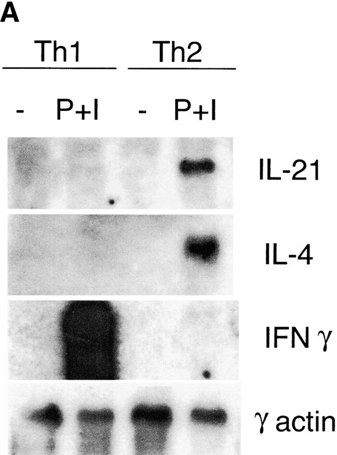
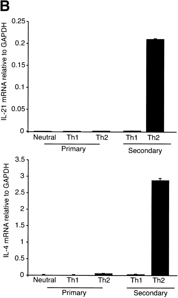
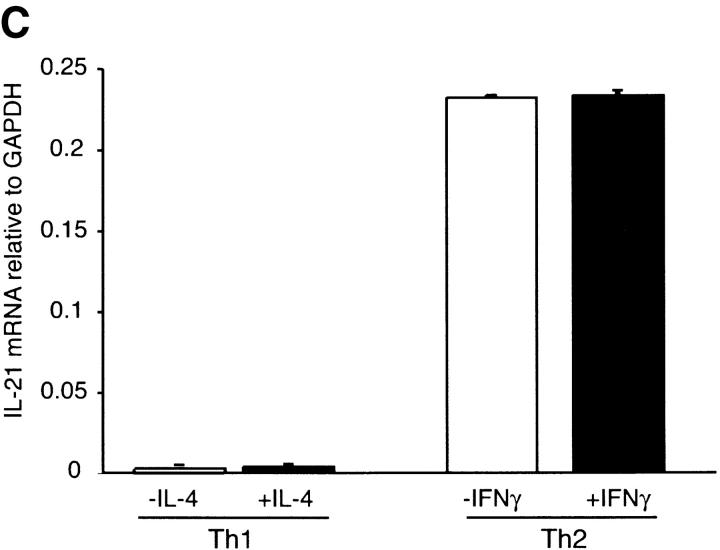
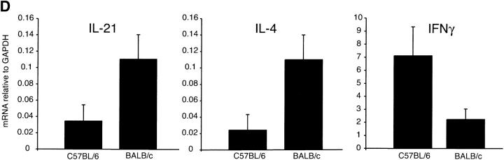
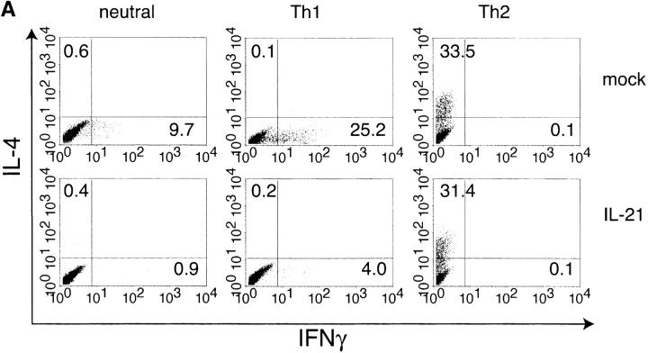
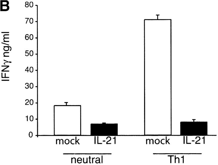
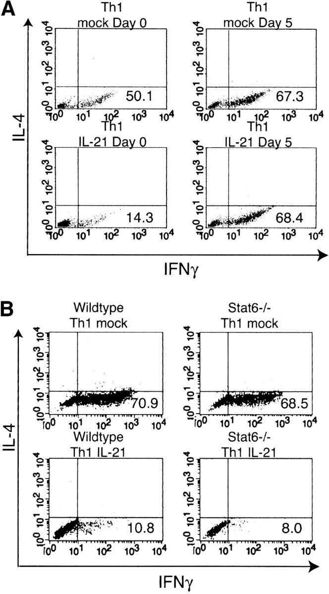
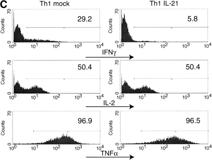
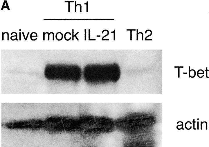
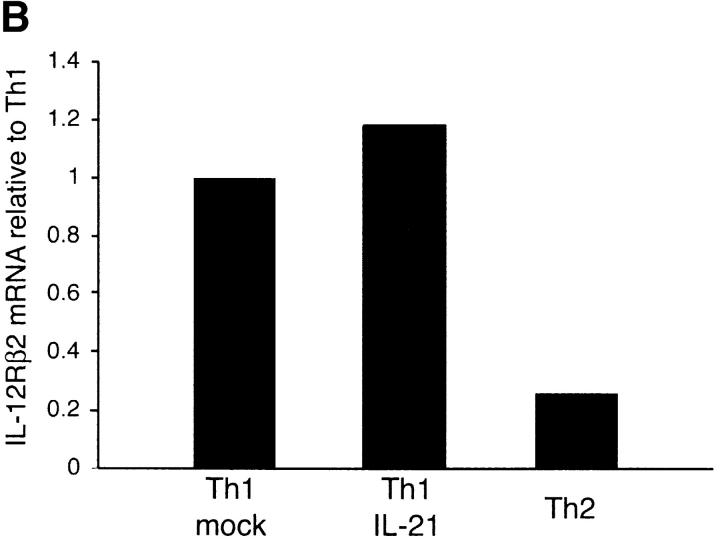
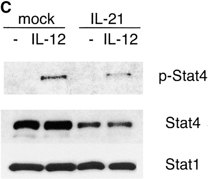
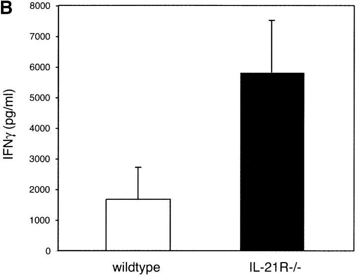
The cytokine potential of developing T helper (Th) cells is directly shaped both positively and negatively by the cytokines expressed by the effector Th cell subsets. Here we find that the recently identified cytokine, interleukin (IL)-21, is preferentially expressed by Th2 cells when compared with Th1 cells generated in vitro and in vivo. Exposure of naive Th precursors to IL-21 inhibits interferon (IFN)-gamma production from developing Th1 cells. The repression of IFN-gamma production is specific in that the expression of other Th1 and Th2 cytokines is unaffected. IL-21 decreases the IL-12 responsiveness of developing Th cells by specifically reducing both signal transducer and activator of transcription 4 protein and mRNA expression. These results suggest that Th2 cell-derived IL-21 regulates the development of IFN-gamma-producing Th1 cells which could serve to amplify a Th2 response.


Figures

Figure 1.
Figure 1.
IL-21 is a Th2 cytokine. (A) Thp cells were cultured under Th1 and Th2 skewing conditions for 6 d. The cells were left resting (−) or restimulated with PMA/Ionomycin (P+I) for 4 h. Similar results were observed for 6 and 24 h after stimulation (unpublished data). RNA was purified and assessed for cytokine expression by Northern blot analysis. The results shown are representative of three independent experiments. (B) Thp cells were cultured under neutral, Th1, and Th2 skewing conditions. RNA was purified 24 h after primary and secondary anti-CD3 stimulation. Cytokine expression was assessed in duplicate by RealTime PCR and shown relative to GAPDH. (C) Thp cells were cultured under Th1 and Th2 skewing conditions for 5 d. IL-4 or IFN-γ were added to indicated cultures 24 h before secondary stimulation with anti-CD3. RNA was purified 24 h after secondary stimulation and IL-21 expression was assessed in duplicate and shown relative to GAPDH by RealTime PCR. (D) Cohorts of eight BALB/c and C57BL/6 mice were infected with L. major in hind footpads. After 6 wk CD4+ T cells from draining lymph nodes were purified and stimulated with anti-CD3. RNA was purified 6 h after stimulation and cytokine expression was assessed relative to GAPDH by RealTime PCR.
Figure 1.
Figure 1.
IL-21 is a Th2 cytokine. (A) Thp cells were cultured under Th1 and Th2 skewing conditions for 6 d. The cells were left resting (−) or restimulated with PMA/Ionomycin (P+I) for 4 h. Similar results were observed for 6 and 24 h after stimulation (unpublished data). RNA was purified and assessed for cytokine expression by Northern blot analysis. The results shown are representative of three independent experiments. (B) Thp cells were cultured under neutral, Th1, and Th2 skewing conditions. RNA was purified 24 h after primary and secondary anti-CD3 stimulation. Cytokine expression was assessed in duplicate by RealTime PCR and shown relative to GAPDH. (C) Thp cells were cultured under Th1 and Th2 skewing conditions for 5 d. IL-4 or IFN-γ were added to indicated cultures 24 h before secondary stimulation with anti-CD3. RNA was purified 24 h after secondary stimulation and IL-21 expression was assessed in duplicate and shown relative to GAPDH by RealTime PCR. (D) Cohorts of eight BALB/c and C57BL/6 mice were infected with L. major in hind footpads. After 6 wk CD4+ T cells from draining lymph nodes were purified and stimulated with anti-CD3. RNA was purified 6 h after stimulation and cytokine expression was assessed relative to GAPDH by RealTime PCR.
Figure 2.
Figure 2.
Impaired IFN-γ production from IL-21–treated Th cells. (A) Thp cells were cultured under neutral, Th1, and Th2 skewing conditions for 1 wk in the presence of 20 ng/ml IL-21 or mock supernatants. Cytokine production was assessed by intracellular cytokine staining 4 h after restimulation with PMA/Ionomycin. Results are representative of at least 10 experiments. (B) Thp cells were cultured in the presence of 20 ng/ml IL-21 or mock supernatants under neutral and Th1 conditions for 48 h. Culture supernatants were assessed for IFN-γ production by ELISA.
Figure 2.
Figure 2.
Impaired IFN-γ production from IL-21–treated Th cells. (A) Thp cells were cultured under neutral, Th1, and Th2 skewing conditions for 1 wk in the presence of 20 ng/ml IL-21 or mock supernatants. Cytokine production was assessed by intracellular cytokine staining 4 h after restimulation with PMA/Ionomycin. Results are representative of at least 10 experiments. (B) Thp cells were cultured in the presence of 20 ng/ml IL-21 or mock supernatants under neutral and Th1 conditions for 48 h. Culture supernatants were assessed for IFN-γ production by ELISA.
Figure 3.
Figure 3.
IL-21 specifically inhibits IFN-γ production from developing Th1 cells. (A) Thp cells were cultured under Th1 skewing conditions. 20 ng/ml IL-21 or mock supernatant was added either at the beginning of culture (day 0) or 24 h before restimulation and analysis (day 5). Cytokine production was assessed by intracellular cytokine staining. (B) Thp cells purified from wild-type or STAT-6–deficient mice were cultured for 1 wk under Th1 skewing conditions in the presence of IL-21 or mock supernatant. Cytokine production was assessed by intracellular cytokine staining. (C) Thp cells were cultured under Th1 skewing conditions in the presence of IL-21 or mock supernatant. Cytokine expression was assessed by intracellular cytokine staining.
Figure 3.
Figure 3.
IL-21 specifically inhibits IFN-γ production from developing Th1 cells. (A) Thp cells were cultured under Th1 skewing conditions. 20 ng/ml IL-21 or mock supernatant was added either at the beginning of culture (day 0) or 24 h before restimulation and analysis (day 5). Cytokine production was assessed by intracellular cytokine staining. (B) Thp cells purified from wild-type or STAT-6–deficient mice were cultured for 1 wk under Th1 skewing conditions in the presence of IL-21 or mock supernatant. Cytokine production was assessed by intracellular cytokine staining. (C) Thp cells were cultured under Th1 skewing conditions in the presence of IL-21 or mock supernatant. Cytokine expression was assessed by intracellular cytokine staining.
Figure 4.
Figure 4.
IL-21 inhibits STAT-4 signaling. (A) Thp cells were cultured under Th1 or Th2 skewing conditions. Protein extracts were harvested at the beginning (naive) and 48 h after culture (Th1 and Th2). During which 20 ng/ml IL-21 or mock supernatants were included in the indicated cultures. T-bet and actin expression were determined by Western blot analysis. (B) Thp cells were cultured under Th1 or Th2 skewing conditions for 1 wk. 20 ng/ml IL-21 or mock supernatant was included in indicated cultures. RNA was harvested 24 h after secondary stimulation with anti-CD3 and assessed for IL-12Rβ2 expression by RealTime PCR. The results are representative of three independent experiments. (C) Thp cells were stimulated with anti-CD3 for 48 h in the presence of 20 ng/ml IL-21 or mock supernatants. The cells were then stimulated for 15 min with 1 ng/ml IL-12. Protein extracts were assessed for phosphorylated STAT-4 (p-STAT-4), STAT-4, and STAT-1 by Western blot analysis. Results are representative of four independent experiments. (D) Thp cells were cultured as in C. RNA was harvested and assessed for STAT-4 expression in duplicateby RealTime PCR and shown relative to GAPDH. Results are representative of three independent experiments.
Figure 4.
Figure 4.
IL-21 inhibits STAT-4 signaling. (A) Thp cells were cultured under Th1 or Th2 skewing conditions. Protein extracts were harvested at the beginning (naive) and 48 h after culture (Th1 and Th2). During which 20 ng/ml IL-21 or mock supernatants were included in the indicated cultures. T-bet and actin expression were determined by Western blot analysis. (B) Thp cells were cultured under Th1 or Th2 skewing conditions for 1 wk. 20 ng/ml IL-21 or mock supernatant was included in indicated cultures. RNA was harvested 24 h after secondary stimulation with anti-CD3 and assessed for IL-12Rβ2 expression by RealTime PCR. The results are representative of three independent experiments. (C) Thp cells were stimulated with anti-CD3 for 48 h in the presence of 20 ng/ml IL-21 or mock supernatants. The cells were then stimulated for 15 min with 1 ng/ml IL-12. Protein extracts were assessed for phosphorylated STAT-4 (p-STAT-4), STAT-4, and STAT-1 by Western blot analysis. Results are representative of four independent experiments. (D) Thp cells were cultured as in C. RNA was harvested and assessed for STAT-4 expression in duplicateby RealTime PCR and shown relative to GAPDH. Results are representative of three independent experiments.
Figure 5.
Figure 5.
Enhanced DTH responses in IL-21R–deficient mice. (A) Specific footpad swelling of wild-type and IL-21R–deficient (IL-21R−/−) mice was determined by subtracting nonspecific swelling in the PBS-injected footpad from the TNP-KLH-induced swelling. Each data point represents one mouse and horizontal lines indicate averages. Results are pooled of two independent experiments. (B) Purified CD4+ T cells from the draining lymph nodes of immunized mice were stimulated in vitro with 250 μg/ml TNP-KLH and irradiated APCs. Supernatants were analyzed for IFN-γ levels by ELISA. Results shown are the average of two mice from each genotype performed in duplicate.
Figure 5.
Figure 5.
Enhanced DTH responses in IL-21R–deficient mice. (A) Specific footpad swelling of wild-type and IL-21R–deficient (IL-21R−/−) mice was determined by subtracting nonspecific swelling in the PBS-injected footpad from the TNP-KLH-induced swelling. Each data point represents one mouse and horizontal lines indicate averages. Results are pooled of two independent experiments. (B) Purified CD4+ T cells from the draining lymph nodes of immunized mice were stimulated in vitro with 250 μg/ml TNP-KLH and irradiated APCs. Supernatants were analyzed for IFN-γ levels by ELISA. Results shown are the average of two mice from each genotype performed in duplicate.

References

    1. Abbas, A.K., K.M. Murphy, and A. Sher. 1996. Functional diversity of helper T lymphocytes. Nature. 383:787–793. - PubMed
    1. Glimcher, L.H., and K.M. Murphy. 2000. Lineage commitment in the immune system: the T helper lymphocyte grows up. Genes Dev. 14:1693–1711. - PubMed
    1. O'Garra, A. 1998. Cytokines induce the development of functionally heterogeneous T helper cell subsets. Immunity. 8:275–283. - PubMed
    1. Maggi, E., P. Parronchi, R. Manetti, C. Simonelli, M.P. Piccinni, F.S. Rugiu, M. De Carli, M. Ricci, and S. Romagnani. 1992. Reciprocal regulatory effects of IFN-γ and IL-4 on the in vitro development of human Th1 and Th2 clones. J. Immunol. 148:2142–2147. - PubMed
    1. Moore, K.W., R. de Waal Malefyt, R.L. Coffman, and A. O'Garra. 2001. Interleukin-10 and the interleukin-10 receptor. Annu. Rev. Immunol. 19:683–765. - PubMed

Publication types