. 2002 Oct 7;196(7):991-8.
doi: 10.1084/jem.20021186.

Virulent but not avirulent Mycobacterium tuberculosis can evade the growth inhibitory action of a T helper 1-dependent, nitric oxide Synthase 2-independent defense in mice

Affiliations

Virulent but not avirulent Mycobacterium tuberculosis can evade the growth inhibitory action of a T helper 1-dependent, nitric oxide Synthase 2-independent defense in mice

Yu-Jin Jung et al. J Exp Med. .

Abstract

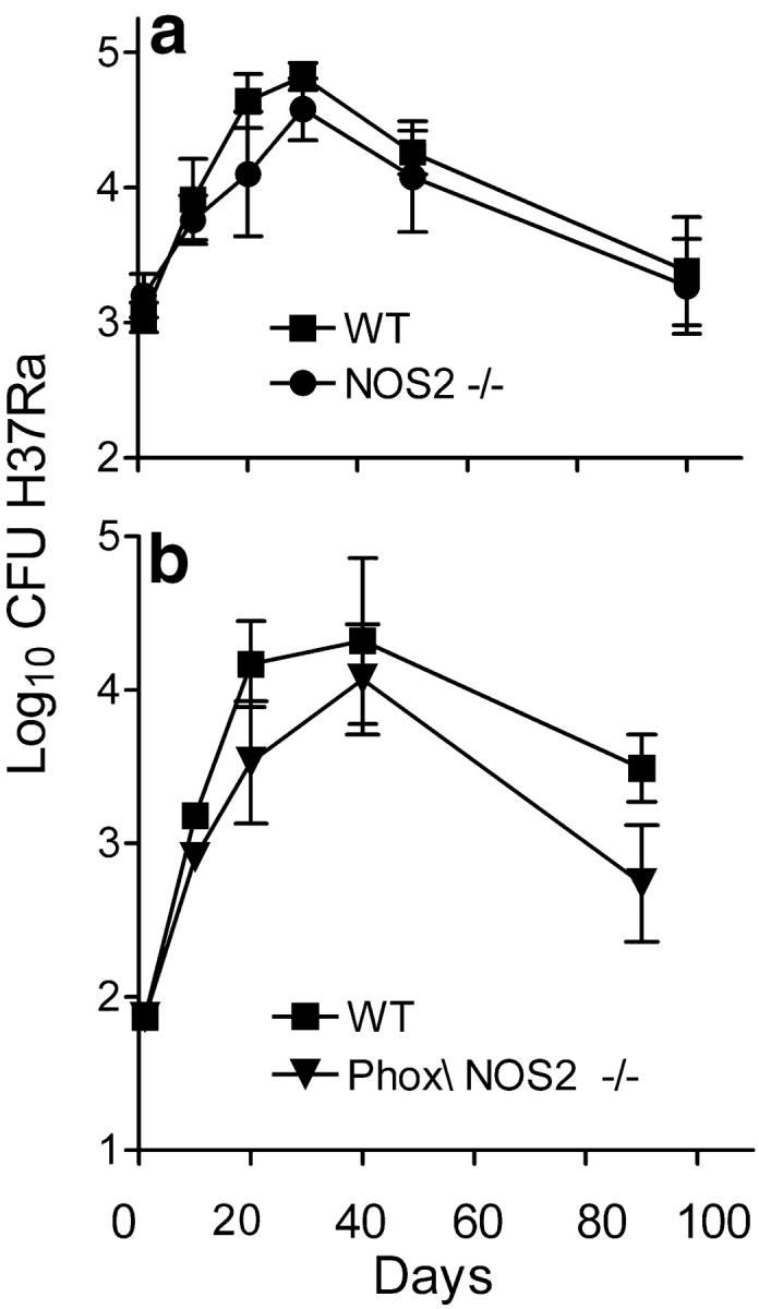
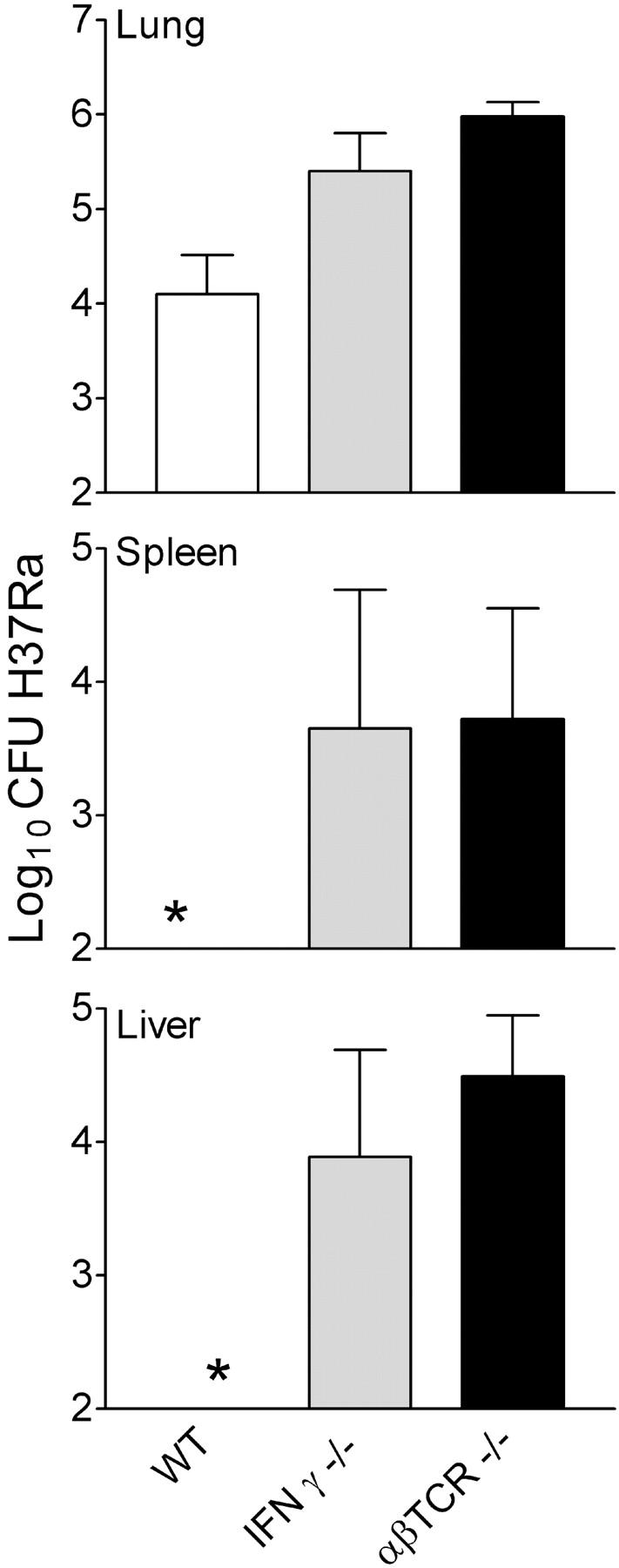
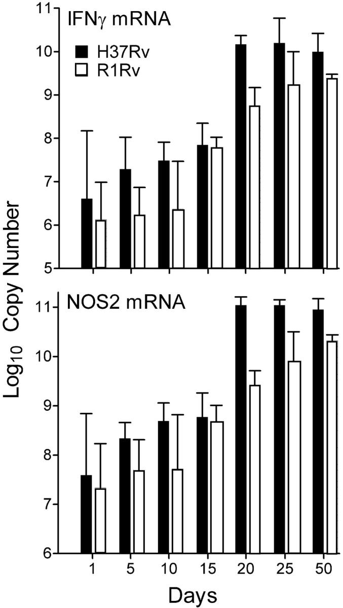
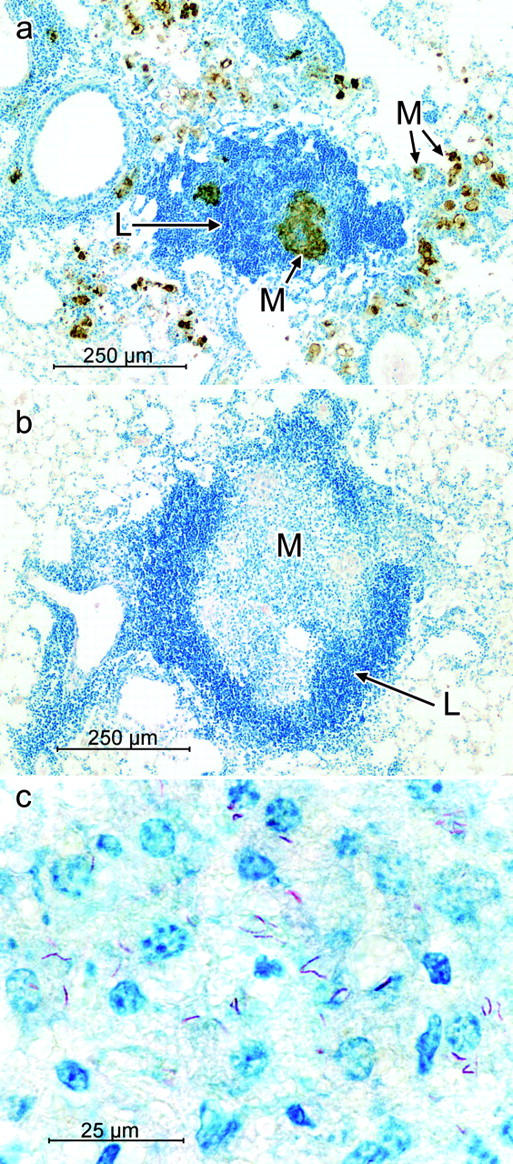
Control of infection with virulent Mycobacterium tuberculosis (Mtb) in mice is dependent on the generation of T helper (Th)1-mediated immunity that serves, via secretion of interferon (IFN)-gamma and other cytokines, to upregulate the antimycobacterial function of macrophages of which the synthesis of inducible nitric oxide synthase (NOS)2 is an essential event. As a means to understanding the basis of Mtb virulence, the ability of gene-deleted mice incapable of making NOS2 (NOS2(-/-)), gp91(Phox) subunit of the respiratory burst NADPH-oxidase complex (Phox(-/-)), or either enzyme (NOS2/Phox(-/-)), to control airborne infection with the avirulent R1Rv and H37Ra strains of Mtb was compared with their ability control infection with the virulent H37Rv strain. NOS2(-/-), Phox(-/-), and NOS2/Phox(-/-) mice showed no deficiency in ability to control infection with either strain of avirulent Mtb. By contrast, NOS2(-/-) mice, but not Phox(-/-) mice, were incapable of controlling H37Rv infection and died early from neutrophil-dominated lung pathology. Control of infection with avirulent, as well as virulent Mtb, depended on the synthesis of IFN-gamma, and was associated with a substantial increase in the synthesis in the lungs of mRNA for IFN-gamma and NOS2, and with production of NOS2 by macrophages at sites of infection. The results indicate that virulent, but not avirulent, Mtb can overcome the growth inhibitory action of a Th1-dependent, NOS2-independent mechanism of defense.


Figures

Figure 1.
Figure 1.
Growth of H37Rv in the lungs, livers, and spleens of WT, Phox−/−, NOS2−/−, and NOS2/Phox−/− mice infected with 102 CFU aerogenically. Whereas in WT and Phox−/− mice growth of H37Rv was controlled from day 20 of infection on, growth of the pathogen was progressive in NOS2−/− and NOS2/Phox−/− mice. Means of five mice per time point ±SD.
Figure 2.
Figure 2.
Growth of R1Rv in lungs, livers, and spleens of WT, Phox−/−, NOS2−/−, and NOS2/Phox−/− mice infected aerogenically with ∼102 CFU. Growth of R1Rv was identical in the lungs of WT and NOS2−/− mice, in that it was controlled at day 20, after which infection slowly resolved. Growth of R1Rv in Phox−/− and NOS2/Phox−/− mice was controlled at about a 1 log lower level than in WT mice after which infection slowly resolved. Dissemination of R1Rv to the liver and spleen was slower in Phox−/− and NOS2/Phox−/− mice, although by day 60 all groups showed similar CFU numbers in these organs. Means of five mice per time point ±SD.
Figure 3.
Figure 3.
Growth of H37Ra in WT and mutant mice. Results of two experiments (a and b) showing that growth H37Ra in WT and NOS2−/− mice (a) was identical, in that in both cases H37Ra grew for 20–30 d in the lung, after which infection slowly resolved. H37Ra also grew similarly in NOS2/Phox−/− mice, except that infection was controlled at a lower level in the latter mice. Means of five mice per time point ±SD.
Figure 4.
Figure 4.
Number of R1Rv in the lungs, livers, and spleens of WT, TCR-αβ−/−, and IFN-γ−/− mice at day 50 of an aerogenic initiated with 102 CFU. Infection was exacerbated substantially in all organs of the mutant mice. Means of five mice ±SD.
Figure 5.
Figure 5.
Number of H37Ra in lungs, livers, and spleens of WT, TCR-αβ−/−, and IFN-γ−/− mice at day 50 of an aerogenic infection initiated with 5 × 10² CFU. Infection in the lungs was significantly higher in mutant mice. H37Ra did not disseminate to the liver and spleen in WT mice, but did so in mutant mice incapable of expressing immunity. Means of five mice ±SD.
Figure 6.
Figure 6.
Results of a real-time RT-PCR study of IFN-γ and NOS2 gene expression in the lungs of WT mice infected aerogenically with 102 CFU of H37Rv or R1Rv. Copy numbers of mRNA for IFN-γ and NOS2 per total lung RNA increase significantly between days 11 and 20 of infection with either organism, and were sustained from day 20 to day 50 when the study was terminated. However, increased IFN-γ and NOS2 mRNA synthesis was significantly lower in the case of lungs infected with R1Rv. Means of three mice per time point ±SD.
Figure 7.
Figure 7.
Micrographs of section of lung from a WT and an NOS2−/− mouse infected 40 d earlier with 102 CFU of H37Rv aerogenically. Lesions in WT lung at low power (a) consisted of aggregates of macrophages stained for NOS2 (brown) in close proximity to aggregates to lymphoid cells (dark blue). At higher power (b) NOS2-stained macrophages were seen to contain acid-fast bacilli (red). In contrast, lesions in NOS2−/− lungs at low power (c) were seen to be composed of large numbers of neutrophils that at higher power (d) were seen to contain acid-fast bacilli and to be in the process of degeneration. No cells in NOS2−/− lungs stained for NOS2. Original magnifications: 80× for a and c; 470× for b and d.
Figure 8.
Figure 8.
Micrographs of lung lesions in WT and NOS2−/− mice infected with 102 CFU of R1Rv aerogenically. At lower power, (a) lesions in WT lungs were composed of aggregates of lymphoid cells (blue) in close association with aggregates of macrophages containing NOS2 (brown). In the lungs of NOS2−/− mice at low power (b) lesions were seen to consist typically of a central core of macrophages surrounded by a mantle of lymphoid cells (blue). At higher power (c) none of the macrophages in the central core stained for NOS2, and some of them were infected with acid-fast bacilli (red). Original magnifications: 92× for a and b: 740× for c.

Similar articles

Cited by

References

    1. Mogues, T., M.E. Goodrich, L. Ryan, R. LaCourse, and R.J. North. 2001. The relative importance of T cell subsets in immunity and immunopathology of airborne Mycobacterium tuberculosis infection in mice. J. Exp. Med. 193:271–380. - PMC - PubMed
    1. Boom, W.H. 1996. The role of T cell subsets in Mycobacterium tuberculosis infection. Infect. Agents Dis. 5:73–81. - PubMed
    1. Cooper, A.M., B.M. Saunders, C.D. D'Souza, A.A. Frank, and I.M. Orme. 1997. Bull Inst. Pasteur. 95:85–95.
    1. Flynn, J.L., and J. Chan. 2001. Immunology of tuberculosis. Annu. Rev. Immunol. 19:93–129. - PubMed
    1. Raupach, B., and S.H. Kaufmann. 2001. Immune response to intracellular bacteria. Curr. Opin. Immunol. 13:417–428. - PubMed

Publication types

MeSH terms

Substances