. 2002 Oct;110(7):933-41.
doi: 10.1172/JCI14268.

Inhibition of endothelial cell survival and angiogenesis by protein kinase A

Affiliations

Inhibition of endothelial cell survival and angiogenesis by protein kinase A

Semi Kim et al. J Clin Invest. 2002 Oct.

Abstract

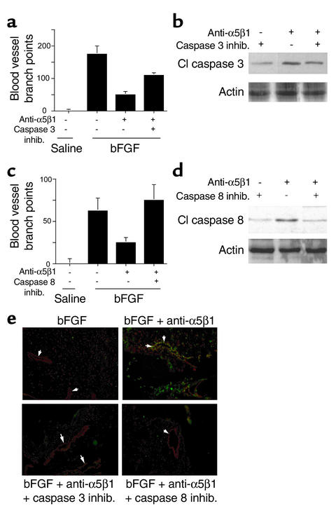
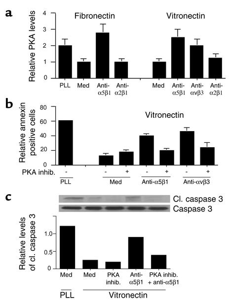
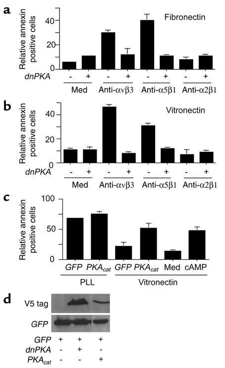
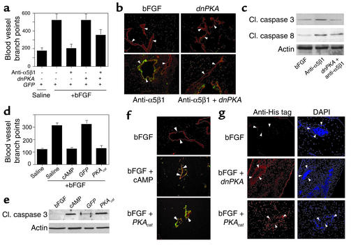
Receptors for the provisional ECM are important regulators of angiogenesis. One of these receptors, integrin alpha5beta1, plays a critical role in tumor- and growth factor-induced angiogenesis, because antagonists of this integrin potently inhibit angiogenesis and tumor growth. Here we show that the integrin alpha5beta1 promotes endothelial cell survival during angiogenesis in vivo by suppressing the activity of protein kinase A (PKA). Antagonists of integrin alpha5beta1 activate PKA, which then leads to the activation of caspase-8 and induction of apoptosis. Direct activation of PKA by cAMP or by expression of the PKA catalytic subunit also induces endothelial cell apoptosis, resulting in angiogenesis inhibition in vivo. Our studies indicate that ligation of integrin alpha5beta1 during angiogenesis suppresses an apoptotic program that is dependent on PKA. These studies also indicate that induction of endothelial cell apoptosis in vivo by genetic or pharmacological activation of PKA may be a useful strategy to inhibit angiogenesis.


Figures

Figure 1
Figure 1
Fibronectin and integrin α5β1 support endothelial cell survival. (ac) HUVECs were maintained in suspension (SUS) or on fibronectin-coated (Fn) or poly-L-lysine–coated (PLL) plates. (a) The percentage of annexin V–positive cells on poly-L-lysine– or fibronectin-coated dishes was determined at regular intervals from 0 to 8 hours. (b) Cell lysates prepared after 4 hours of attachment were immunoblotted to detect intact (116 kDa) and cleaved PARP (85 kDa). The ratio of intact to cleaved PARP was determined by densitometry. Cl., cleaved. (c) Soluble DNA extracted from cells attached to poly-L-lysine or fibronectin was electrophoresed on 1.6% agarose gels. Relative DNA cleavage was determined by densitometry. (df) HUVECs were plated on fibronectin, anti-α5β1, or control Ab-coated plates. (d) The percentage of annexin V–positive cells was determined from 0 to 8 hours. (e) Cell lysates were immunoblotted to detect intact and cleaved PARP. (f) DNA fragmentation was evaluated as in c. cIgG, control immunoglobulin.
Figure 2
Figure 2
Integrin α5β1 supports endothelial cell survival during angiogenesis in vivo. (a) CAMs were stimulated with bFGF or saline and then treated for 24 hours with saline, anti-α5β1, and control Ab’s. CAMs were then injected with 50 μl FITC-annexin V, harvested 2 hours later, and analyzed directly by confocal microscopy. (b) Green pixels (annexin V positive) present per optical section were quantified. (c) CAMs treated as in a were cryosectioned and immunostained with anti–cleaved caspase-3 (anti-cl.csp3) (green) and anti-vWF (red). Cleaved caspase-3–positive blood vessels are yellow (arrows). (d) CAMs treated as in a were cryosectioned and immunostained with anti-vWF (red) and for DNA fragmentation (TUNEL staining, green). Arrows indicate blood vessels. Yellow structures are apoptotic blood vessels. (e) Soluble DNA isolated from CAMs treated as in a was electrophoresed on 1.6% agarose gels. Molecular-weight markers are 1-kb DNA ladders. Relative DNA cleavage was determined by densitometry. (f) Individual cells isolated from CAMs treated as in a were stained with FITC-annexin V.
Figure 3
Figure 3
Unligated integrin α5β1 regulates endothelial cell survival. HUVECs were plated on poly-L-lysine–coated, fibronectin-coated (a and b), or vitronectin-coated (c and d) culture plates in culture medium (med) or culture medium containing anti-α5β1, anti-αvβ3, anti-α2β1 Ab’s. After 1 hour, cell attachment was determined (b and d). After 24 hours, the percentage of FITC-annexin V–positive (a and c) cells was determined. (e) HUVECs plated on vitronectin-coated plates in the presence of function-blocking anti-α5β1 or control Ab’s were collected at regular intervals from 0 to 8 hours and PARP cleavage assessed by Western blotting. Relative PARP cleavage levels were determined by densitometry.
Figure 4
Figure 4
Unligated integrin α5β1 induces caspase-3 and -8, but not -9, activation. HUVECs were plated on (a) fibronectin-, (b) vitronectin-, or (a and b) poly-L-lysine–coated culture plates in the presence of anti-α5β1 Ab’s and 50 μM z-DEVD-fmk (caspase-3) or z-IETD-fmk (caspase-8) inhibitors or vehicle control (0.33% DMSO) for 24 hours. The percentage of annexin V–positive cells was then determined. inhib, inhibitor. (c and d) Caspase-3 and -8 activities were determined in HUVECs plated on vitronectin-coated or poly-L-lysine–coated plates in the presence of culture medium, anti-α5β1, or control Ab’s. (e) Cell lysates were immunoblotted with anti–caspase-3 and anti–cleaved caspase-3 Ab’s. Relative caspase-3 cleavage was determined by densitometry. (f) Cell lysates were immunoblotted with anti–caspase-9 and anti–cleaved caspase-9 Ab’s.
Figure 5
Figure 5
Integrin antagonists induce caspase-3– and -8–dependent apoptosis in vivo. (ad) CAMs stimulated with saline or bFGF were treated with 2.5% DMSO (vehicle control), anti-α5β1, or anti-α5β1 with 500 μM caspase-3 (a and b) or caspase-8 (c and d) inhibitors. (a and c) Blood vessel branch points were quantified after 48 hours. (b and d) Caspase-3 and -8 cleavage was evaluated by Western blotting with (b) anti–cleaved caspase-3 or (d) anti–cleaved caspase-8 Ab’s and anti-actin Ab’s. (e) Cryosections of CAMs treated as in ad were immunostained for vWF expression (red) and to detect DNA fragmentation (TUNEL staining, green). Arrows indicate blood vessels. Apoptotic vessels appear yellow.
Figure 6
Figure 6
Unligated α5β1-mediated death is PKA dependent. (a) PKA activity was measured in HUVECs attached to poly-L-lysine, fibronectin, or vitronectin in the presence or absence of integrin antagonists. (b) HUVECs were plated on vitronectin-coated or poly-L-lysine–coated culture plates in the presence or absence of anti-α5β1 or anti-αvβ3 Ab’s, a selective PKA inhibitor (1 μM HA1004), or anti-integrin Ab’s in combination with 1 μM HA1004. After 24 hours, the percentage of FITC-annexin–positive cells was determined. (c) Cell lysates from b were immunoblotted with anti–caspase-3 and anti–cleaved caspase-3 Ab’s. Relative caspase-3 cleavage was determined by densitometry.
Figure 7
Figure 7
PKA negatively regulates cell survival. (a and b) HUVECs transfected with GFP (–) or a dnPKA (+) were plated on (a) fibronectin-coated, (b) vitronectin-coated, or poly-L-lysine–coated plates in the absence or presence of anti-α5β1, anti-αvβ3, or anti-α2β1. After 24 hours, the percentage of annexin V–positive cells was determined. (c) HUVECs treated with culture medium or dibutyryl cAMP (250 μM) and HUVECs transfected with GFP or the catalytic subunit of PKA (PKAcat) were plated on vitronectin-, or poly-L-lysine–coated plates. After 24 hours, the percentage of annexin V–positive cells was determined. (d) Expression of transgenes was detected by Western blotting cell lysates with anti-GFP or anti-V5.
Figure 8
Figure 8
PKA inhibits angiogenesis by inducing apoptosis. (a) CAMs stimulated with bFGF were transfected 24 hours later by placing 4 μg pcDNA/V5/His dnPKA or N1-GFP expression plasmid on CAMs. CAMs were treated on the same day with saline or anti-α5β1 Ab’s and were harvested 48 hours later. Blood vessel branch points were quantified. (b) Cryosections of CAMs from a were immunostained with anti-vWF (red) and were stained to detect fragmented DNA by the TUNEL method (green). Arrows indicate blood vessels. Apoptotic blood vessels appear yellow. (c) Western blots of lysates prepared from CAMs treated as in a were immunoblotted with anti–cleaved caspase-3 and anti–cleaved caspase –8, as well as anti-actin as a loading control. (d) CAMs stimulated with bFGF were treated with saline or 250 μM cAMP or were transfected by placing 4 μg pcDNA/V5/His PKAcat or N1-GFP expression plasmid on stimulated CAMs. Blood vessel branch points were quantified 48 hours later. (e) Cryosections of CAMs from d were immunostained with anti-vWF (red) and were stained to detect fragmented DNA by the TUNEL method (green). Arrows indicate blood vessels. Apoptotic blood vessels appear yellow. (f) Detergent lysates prepared from freshly excised CAMs from d were immunoblotted for expression of cleaved caspase-3 and actin as a loading control. (g) Cryosections of CAMs from a and d were immunostained with anti-pentaHis (red) to detect expression of His-tagged transgenes in the transfected CAM tissue. Sections were counterstained with DAPI. Arrows indicate blood vessels.

Comment in

References

    1. Varner JA. The role of vascular cell integrin αvβ3 and αvβ5 in angiogenesis. EXS. 1997;79:361–390. - PubMed
    1. Asahara T, et al. VEGF contributes to postnatal neovascularization by mobilizing bone marrow derived endothelial progenitor cells. EMBO J. 1999;18:3964–3972. - PMC - PubMed
    1. Meredith JE, Jr, Fazeli B, Schwartz MA. The ECM as a cell survival factor. Mol Biol Cell. 1993;4:953–961. - PMC - PubMed
    1. Brooks PC, et al. Integrin αvβ3 antagonists promote tumor regression by inducing apoptosis of angiogenic blood vessels. Cell. 1994;79:1157–1164. - PubMed
    1. Boudreau N, Sympson CJ, Werb Z, Bissell M. Suppression of ICE and apoptosis in mammary epithelial cells by ECM. Science. 1995;267:891–893. - PMC - PubMed

Publication types