. 2002 Nov;22(21):7491-500.
doi: 10.1128/MCB.22.21.7491-7500.2002.

Identification, cDNA cloning, and targeted deletion of p70, a novel, ubiquitously expressed SH3 domain-containing protein

Affiliations

Identification, cDNA cloning, and targeted deletion of p70, a novel, ubiquitously expressed SH3 domain-containing protein

Nick Carpino et al. Mol Cell Biol. 2002 Nov.

Abstract

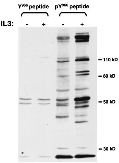
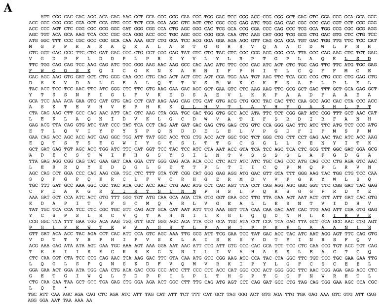
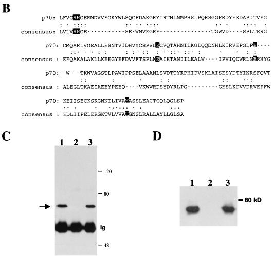
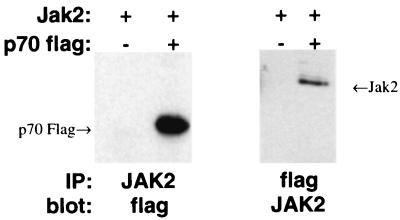
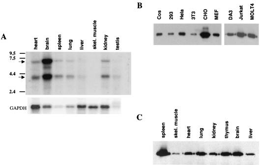
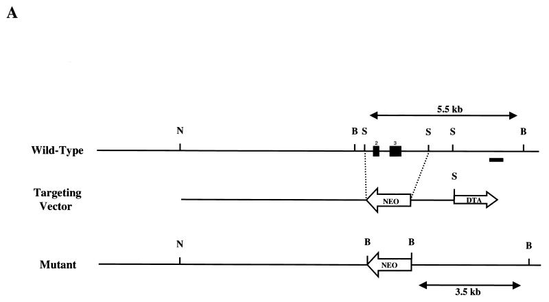
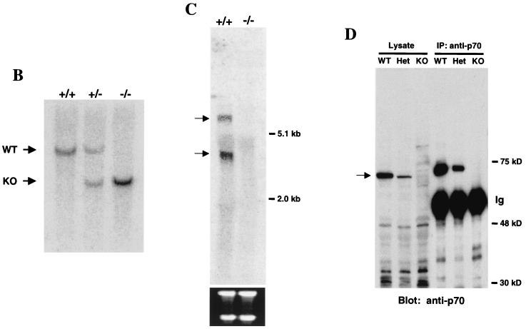
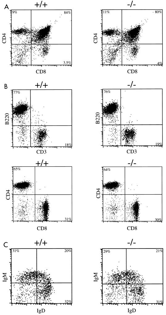
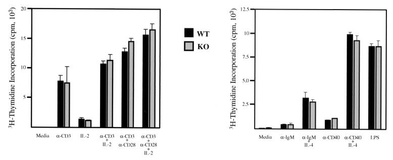
In a screen for proteins that interact with Jak2, we identified a previously uncharacterized 70-kDa protein and cloned the corresponding cDNA. The predicated sequence indicates that p70 contains an SH3 domain and a C-terminal domain with similarities to the catalytic motif of phosphoglycerate mutase. p70 transcripts were found in all tissues examined. Similarly, when an antibody raised against a C-terminal peptide to analyze p70 protein expression was used, all murine tissues examined were found to express p70. To investigate the in vivo role of p70, we generated a p70-deficient mouse strain. Mice lacking p70 are viable, develop normally, and do not display any obvious abnormalities. No differences were detected in various hematological parameters, including bone marrow colony-forming ability, in response to cytokines that utilize Jak2. In addition, no impairment in B- and T-cell development and proliferative ability was detected.


Figures

FIG. 1.
FIG. 1.
Peptide pulldown assay with Jak2-derived peptides. Cells were starved of growth factor overnight and were either left untreated (−) or treated with IL-3 for 5 min (+). Lysates were prepared and incubated with Sepharose beads to which nonphosphorylated Y966 peptide (TSQICKGMEYLGTKR) or tyrosine phosphorylated pY966 peptide (TSQICKGMEpYLGTKR) was conjugated, as outlined in Materials and Methods. Bound proteins were separated by SDS-PAGE, and tyrosine-phosphorylated proteins were analyzed by Western blotting with antiphosphotyrosine antibodies.
FIG. 2.
FIG. 2.
The p70 gene. (A) Nucleotide sequence and predicted amino acid sequence of p70 open reading frame. Underlined amino acids indicate peptides obtained from microsequencing-purified p70 protein, as described in Materials and Methods. (B) Alignment of C-terminal region of p70 with a consensus sequence built by the Protein Family (Pfam) database for members of the phosphoglycerate mutase family (1). The two arginines, two histidines, and glutamic acid predicted to be involved in catalytic activity are highlighted. Identical residues are indicated with two dots; chemically similar residues are indicated with one dot. (C) Peptide competition analysis. Proteins precipitated from lysates of DA3 cells by anti-p70 peptide antibody were separated by SDS-PAGE and transferred to nitrocellulose. The membrane was probed with anti-p70 peptide antibody. Prior to immunoprecipitation, lysates were incubated for 15 min with the following: lane 1, no peptide; lane 2, antigenic peptide; and lane 3, unrelated peptide. Number of kilodaltons is given on right. (D) Interaction of p70 with phosphorylated Jak2 Y966 peptide. DA3 cytoplasmic lysates were subjected to immunoprecipitation with anti-p70 peptide antibodies (lane 1) or affinity pulldown assays utilizing nonphosphorylated Y966 peptide conjugated to Sepharose (lane 2) or phosphorylated Y966 peptide conjugated to Sepharose (lane 3). Blotting antibody was anti-p70 peptide antibody.
FIG. 2.
FIG. 2.
The p70 gene. (A) Nucleotide sequence and predicted amino acid sequence of p70 open reading frame. Underlined amino acids indicate peptides obtained from microsequencing-purified p70 protein, as described in Materials and Methods. (B) Alignment of C-terminal region of p70 with a consensus sequence built by the Protein Family (Pfam) database for members of the phosphoglycerate mutase family (1). The two arginines, two histidines, and glutamic acid predicted to be involved in catalytic activity are highlighted. Identical residues are indicated with two dots; chemically similar residues are indicated with one dot. (C) Peptide competition analysis. Proteins precipitated from lysates of DA3 cells by anti-p70 peptide antibody were separated by SDS-PAGE and transferred to nitrocellulose. The membrane was probed with anti-p70 peptide antibody. Prior to immunoprecipitation, lysates were incubated for 15 min with the following: lane 1, no peptide; lane 2, antigenic peptide; and lane 3, unrelated peptide. Number of kilodaltons is given on right. (D) Interaction of p70 with phosphorylated Jak2 Y966 peptide. DA3 cytoplasmic lysates were subjected to immunoprecipitation with anti-p70 peptide antibodies (lane 1) or affinity pulldown assays utilizing nonphosphorylated Y966 peptide conjugated to Sepharose (lane 2) or phosphorylated Y966 peptide conjugated to Sepharose (lane 3). Blotting antibody was anti-p70 peptide antibody.
FIG. 3.
FIG. 3.
Coimmunoprecipitation of Jak2 and p70. Cos cells were cotransfected as indicated, and lysates were subjected to immunoprecipitation (IP)/Western blot analysis with the indicated antibody.
FIG. 4.
FIG. 4.
p70 expression analysis. (A) Northern blots containing poly(A)+ RNA from multiple murine tissues were probed with a p70 cDNA probe (upper panel) or a glyceraldehyde-3-phosphate dehydrogenase (GAPDH) probe (lower panel). Positions of p70 mRNA and GAPDH mRNA are indicated, as are positions of standard markers, in kilobases. (B) Lysates prepared from the indicated cell lines were subjected to immunoprecipitation/Western blot analysis utilizing anti-p70 peptide antibody. (C) Lysates prepared from the indicated murine tissues were subjected to immunoprecipitation/Western blot analysis utilizing anti-p70 peptide antibody.
FIG. 5.
FIG. 5.
p70 targeting. (A) Structure of the p70 targeting construct. The two filled boxes represent putative exons 2 and 3. NheI (N), BamHI (B), and SphI (S) sites are indicated, as is the location of the probe utilized in Southern analysis to distinguish wild-type and targeted alleles (black bar). Neo, neomycin resistance cassette; DTA, diphtheria toxin cassette. (B) Southern blot of BamHI-digested genomic DNA from +/+, +/−, and −/− mice probed with the external probe illustrated in Fig. 4A. Bands generated by the wild-type (+/+), heterozygote (+/−), and targeted (−/−) alleles are indicated. WT, wild type; KO, knockout. (C) Northern analysis of RNA isolated from wild-type (+/+) or p70-deficient (−/−) thymi. The blot was probed with a p70 cDNA probe that was 3′ to the targeted exons as indicated in Materials and Methods. The bottom panel indicates the 28S and 18S RNAs as loading controls. (D) Immunoprecipitation (IP)/Western blot analysis of levels of p70 protein in splenic lysates prepared from +/+ (WT), +/− (Het), and −/− (KO) littermates.
FIG. 5.
FIG. 5.
p70 targeting. (A) Structure of the p70 targeting construct. The two filled boxes represent putative exons 2 and 3. NheI (N), BamHI (B), and SphI (S) sites are indicated, as is the location of the probe utilized in Southern analysis to distinguish wild-type and targeted alleles (black bar). Neo, neomycin resistance cassette; DTA, diphtheria toxin cassette. (B) Southern blot of BamHI-digested genomic DNA from +/+, +/−, and −/− mice probed with the external probe illustrated in Fig. 4A. Bands generated by the wild-type (+/+), heterozygote (+/−), and targeted (−/−) alleles are indicated. WT, wild type; KO, knockout. (C) Northern analysis of RNA isolated from wild-type (+/+) or p70-deficient (−/−) thymi. The blot was probed with a p70 cDNA probe that was 3′ to the targeted exons as indicated in Materials and Methods. The bottom panel indicates the 28S and 18S RNAs as loading controls. (D) Immunoprecipitation (IP)/Western blot analysis of levels of p70 protein in splenic lysates prepared from +/+ (WT), +/− (Het), and −/− (KO) littermates.
FIG. 6.
FIG. 6.
Flow cytometry analysis of T and B cells. Calculated percentages are displayed within each quadrant. (A) Thymocytes from wild-type and p70-deficient mice were stained with Cyc-conjugated anti-CD4 and FITC-conjugated anti-CD8. (B) Splenocytes from wild-type and p70-deficient mice were stained with Cyc-conjugated anti-B220 and FITC-conjugated anti-CD3 antibodies (top pair). Alternatively, splenocytes were stained with PE-conjugated anti-Thy1.2, Cyc-conjugated anti-CD4, and FITC-conjugated anti-CD8. The CD4/CD8 profile of splenocytes gated on Thy 1.2-positive cells is displayed (bottom panel). (C) Splenocytes from +/+ or −/− mice were stained with FITC-conjugated anti-IgD, PE-conjugated anti-IgM, and Cyc-conjugated anti-B220. The IgM/IgD profile of splenocytes gated on B220-positive cells is displayed. Shown are representative FACS analyses of at least six mice of each genotype.
FIG. 7.
FIG. 7.
T- and B-cell proliferation assays. FACS-purified splenic T cells (left) or FACS-purified splenic B cells (right) were cultured in the presence of the indicated stimuli for 48 h. Cell proliferation was measured by adding 1 μCi of [3H]thymidine to the cultures. Standard deviations for triplicate samples are indicated by the error bars. These data are representative of three T-cell and B-cell proliferation assays. LPS, lipopolysaccharide; WT, wild type; KO, knockout.

References

    1. Bateman, A., E. Birney, L. Cerruti, R. Durbin, L. Etwiller, S. R. Eddy, S. Griffiths-Jones, K. L. Howe, M. Marshall, and E. L. Sonnhammer. 2002. The Pfam protein families database. Nucleic Acids Res. 30:276-280. - PMC - PubMed
    1. Bazan, J. F., R. J. Fletterick, and S. J. Pilkis. 1989. Evolution of a bifunctional enzyme: 6-phosphofructo-2-kinase/fructose-2,6-bisphosphatase. Proc. Natl. Acad. Sci. USA 86:9642-9646. - PMC - PubMed
    1. Capecchi, M. R. 1994. Targeted gene replacement Sci. Am. 270:52-59. - PubMed
    1. Carter-Su, C., L. Rui, and M. R. Stofega. 2000. SH2-B and SIRP: JAK2 binding proteins that modulate the actions of growth hormone. Recent Prog. Horm. Res. 55:293-311. - PubMed
    1. Damen, J. E., L. Liu, H. Wakao, A. Miyajima, P. Rosten, A. B. Jefferson, P. W. Majerus, J. Krosl, R. K. Humphries, and G. Krystal. 1997. The role of erythropoietin receptor tyrosine phosphorylation in erythropoietin-induced proliferation. Leukemia 3(Suppl.):423-425. - PubMed

Publication types

MeSH terms