. 2002 Nov;22(21):7603-13.
doi: 10.1128/MCB.22.21.7603-7613.2002.

Targeted disruption of NFATc3, but not NFATc4, reveals an intrinsic defect in calcineurin-mediated cardiac hypertrophic growth

Affiliations

Targeted disruption of NFATc3, but not NFATc4, reveals an intrinsic defect in calcineurin-mediated cardiac hypertrophic growth

Benjamin J Wilkins et al. Mol Cell Biol. 2002 Nov.

Abstract

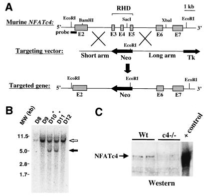
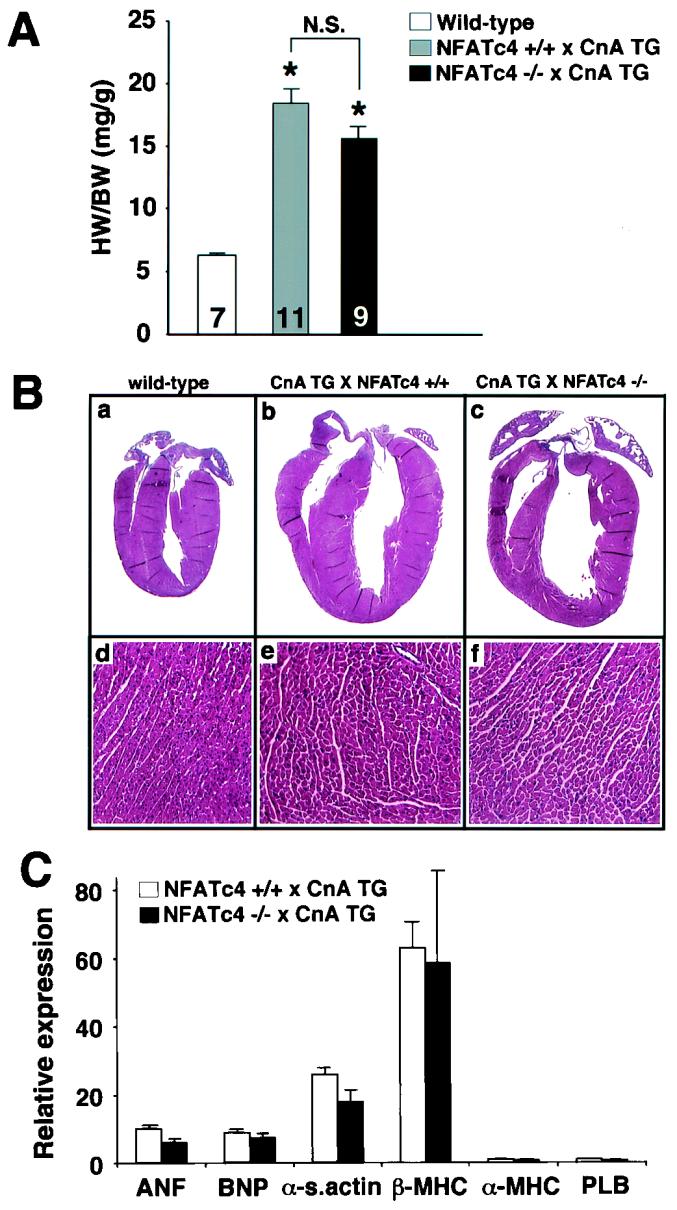
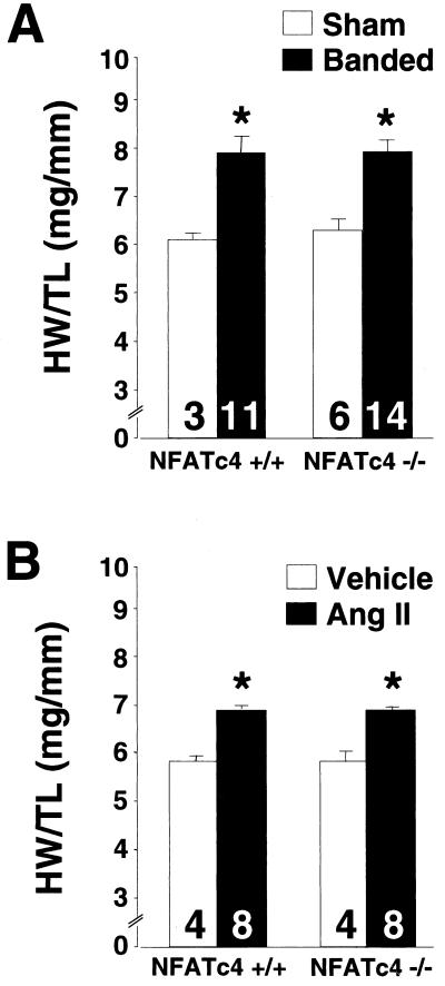
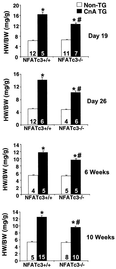
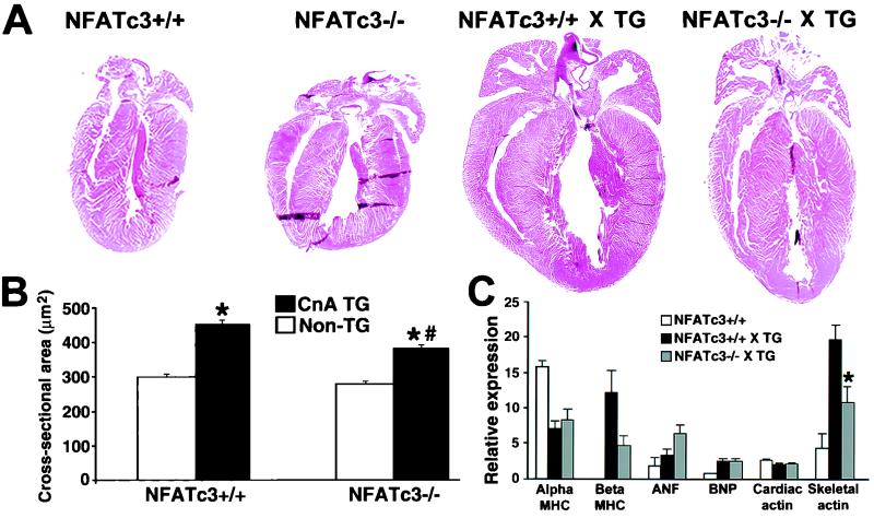
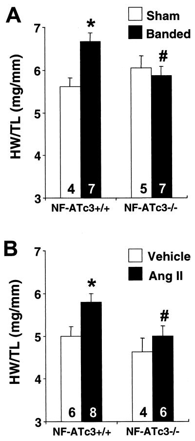
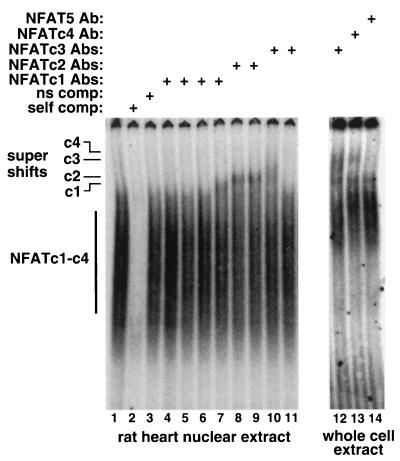
A calcineurin-nuclear factor of activated T cells (NFAT) regulatory pathway has been implicated in the control of cardiac hypertrophy, suggesting one mechanism whereby alterations in intracellular calcium handling are linked to the expression of hypertrophy-associated genes. Although recent studies have demonstrated a necessary role for calcineurin as a mediator of cardiac hypertrophy, the potential involvement of NFAT transcription factors as downstream effectors of calcineurin signaling has not been evaluated. Accordingly, mice with targeted disruptions in NFATc3 and NFATc4 genes were characterized. Whereas the loss of NFATc4 did not compromise the ability of the myocardium to undergo hypertrophic growth, NFATc3-null mice demonstrated a significant reduction in calcineurin transgene-induced cardiac hypertrophy at 19 days, 26 days, 6 weeks, 8 weeks, and 10 weeks of age. NFATc3-null mice also demonstrated attenuated pressure overload- and angiotensin II-induced cardiac hypertrophy. These results provide genetic evidence that calcineurin-regulated responses require NFAT effectors in vivo.


Figures

FIG. 1.
FIG. 1.
Targeted disruption of the murine NFATc4 gene. (A) Genomic structure of the NFATc4 gene, the targeting vector, and the theoretical targeted allele which deletes the Rel homology domain (RHD) of NFATc4. (B) EcoRI-digested genomic Southern blot analysis of targeted ES cell clones D10 and D11 shows an endogenous 10-kb fragment (open arrow) and a targeted 5-kb fragment (solid arrow). (C) NFATc4 Western blot from wild-type and NFATc4−/− adult hearts. A positive control for NFATc4 protein migration was generated by the transfection of Cos cells.
FIG. 2.
FIG. 2.
Gravimetrical, histochemical, and molecular analysis of calcineurin-transgenic mice crossed into the NFATc4-null background. (A) Heart weight/body weight ratios of 18-day-old wild-type, CnTG NFATc4+/+, and CnTG NFATc4−/− mice. The number of mice analyzed in each group is shown within each bar. (B) Representative gross and microscopic (magnification, ×140) histologic heart sections stained with hematoxylin and eosin. (C) Induction of hypertrophy-associated mRNA transcripts in CnTG NFATc4+/+ and CnTG NFATc4−/− mice as measured by RNA dot blot quantitation (i.e., the fold increase over wild-type normalized to GAPDH; n = 4 for all groups). HW, heart weight; BW, body weight; CnA TG, calcineurin transgenic.
FIG. 3.
FIG. 3.
Heart weight/tibia length ratios of 8- to 12-week-old NFATc4−/− and NFATc4+/+ mice subjected to 14 days of abdominal aortic constriction (A) or angiotensin II infusion (432 μg kg−1 day−1) (B). (∗P < 0.05 versus sham- or vehicle-treated controls). The number of mice analyzed in each group is shown within each bar. HW, heart weight; TL, tibia length.
FIG. 4.
FIG. 4.
Heart weight/body weight ratios of NFATc3+/+ and NFATc3−/− mice in the absence or presence of a heart-restricted activated calcineurin transgene at 19 days, 26 days, 6 weeks, and 10 weeks of age. ✽, P < 0.05 versus NFATc3+/+ mice; #, P < 0.05 versus CnTG NFATc3+/+ mice. The number of mice analyzed in each group is shown within each bar. HW, heart weight; BW, body weight; CnA TG, calcineurin transgenic.
FIG. 5.
FIG. 5.
(A) Representative hematoxylin-and-eosin-stained gross histology from 6-week-old mice crossed with the heart-restricted calcineurin transgene. (B) Myofibrillar cross-sectional areas measured by wheat germ agglutinin-TRITC (tetramethyl rhodamine isothiocyanate) staining of histologic heart sections as previously described (27) (n = 250 fibers in each group). ✽, P < 0.05 versus nontransgenic mice; #, P < 0.05 versus CnTG NFATc3+/+ mice. (C) Quantitation of RNA dot blot analysis of hypertrophy-associated transcripts from wild-type (n = 3), CnTG NFATc3+/+ (n = 5), and CnTG NFATc3−/− (n = 4) mice (i.e., the fold increase versus GAPDH × 10). ✽, P < 0.05 versus CnTG NFATc3+/+ mice.
FIG. 6.
FIG. 6.
Heart weight/tibia length ratios of wild-type and NFATc3−/− mice subjected to 14 days of abdominal aortic constriction (A) or angiotensin II infusion (432 μg kg−1 day−1) (B). ✽, P < 0.05 versus NFATc3+/+ mice untreated; #, P < 0.05 versus NFATc3+/+ mice treated. The number of mice analyzed in each group is shown within each bar. HW, heart weight; TL, tibia length.
FIG. 7.
FIG. 7.
Semiquantitative RT-PCR analysis of NFATc1 to -c4 mRNA levels from wild-type hearts (n = 4), NFATc4−/− hearts (n = 3), and NFATc3−/− hearts (n = 3) at 6 weeks of age.
FIG. 8.
FIG. 8.
Western blots of NFAT protein expression in the heart. (A) Nuclear protein extracts from adult human hearts were subjected to Western blotting for NFATc1 to -c4. The positive control consisted of Cos cells transfected with an expression vector for NFATc2 to -c4 or thymocyte extract for NFATc1. (B) Western blot for NFAT5 from human cytoplasmic or nuclear extracts. (C) Protein extracts from wild-type hearts (pool of seven) or α-MHC-CnA-transgenic hearts (pool of six) were Western blotted for NFATc3. The various phosphorylation states are designated by horizontal lines. Myosin, myosin protein that cross-reacts with the NFATc3 primary antibody from the crude mouse heart protein extracts.
FIG. 9.
FIG. 9.
EMSA for NFAT protein expression in the heart. Lanes 1 to 11 consisted of rat heart nuclear protein extracts that were incubated with a consensus NFAT binding site from the IL-4 promoter. Lanes 12 to 14 consisted of protein extracts from cultured neonatal rat cardiomyocytes. Although not all antibodies employed were capable of “supershifting” the NFAT-DNA complexes, certain antibodies successfully shifted NFATc1 to -c4 but not NFAT5.

References

    1. Antos, C. L., T. A. McKinsey, N. Frey, W. Kutschke, J. McAnally, J. M. Shelton, J. A. Richardson, J. A. Hill, and E. N. Olson. 2002. Activated glycogen synthase-3β suppresses cardiac hypertrophy in vivo. Proc. Natl. Acad. Sci. USA 99:907-912. - PMC - PubMed
    1. Beals, C. R., C. M. Sheridan, C. W. Turck, P. Gardner, and G. R. Crabtree. 1997. Nuclear export of NF-ATc enhanced by glycogen synthase kinase-3. Science 275:1930-1934. - PubMed
    1. Blaeser, F., H. Ho, R. Prywes, and T. A. Chatila. 2000. Ca2+-dependent gene expression mediated by MEF2 transcription factors. J. Biol. Chem. 275:197-209. - PubMed
    1. Bueno, O. F., B. J. Wilkins, K. M. Tymitz, B. J. Glascock, T. F. Kimball, J. N. Lorenz, and J. D. Molkentin. 2002. Impaired cardiac hypertrophic response in calcineurin Aβ-deficient mice. Proc. Natl. Acad. Sci. USA 99:4586-4591. - PMC - PubMed
    1. Crabtree, G. R. 1999. Generic signals and specific outcomes: signaling through Ca2+, calcineurin, and NF-AT. Cell 96:611-614. - PubMed

Publication types

MeSH terms