. 2002 Nov;22(21):7622-32.
doi: 10.1128/MCB.22.21.7622-7632.2002.

Intralysosomal cystine accumulation in mice lacking cystinosin, the protein defective in cystinosis

Affiliations

Intralysosomal cystine accumulation in mice lacking cystinosin, the protein defective in cystinosis

Stéphanie Cherqui et al. Mol Cell Biol. 2002 Nov.

Abstract

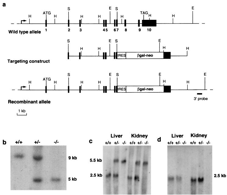
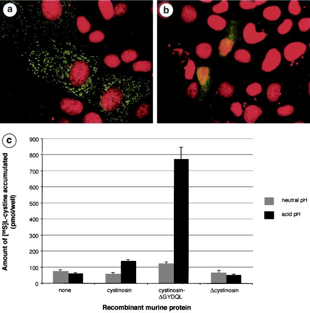
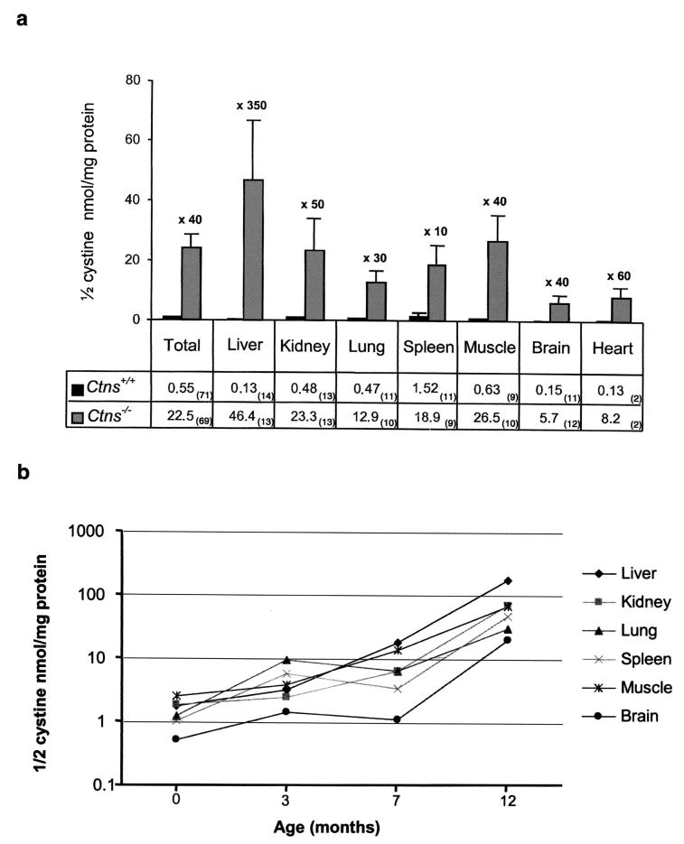
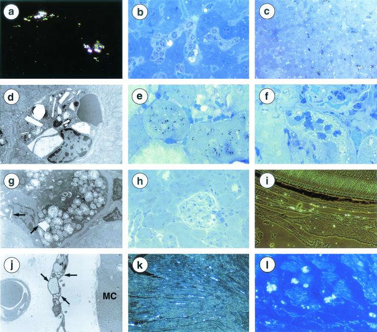
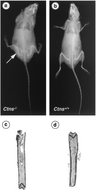
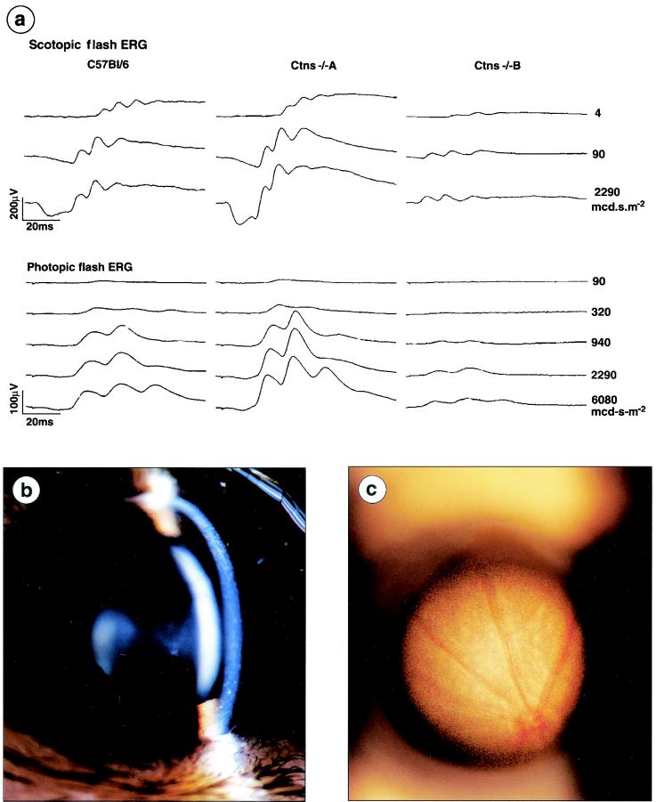
Cystinosis is an autosomal recessive disorder characterized by an accumulation of intralysosomal cystine. The causative gene, CTNS, encodes cystinosin, a seven-transmembrane-domain protein, which we recently showed to be a lysosomal cystine transporter. The most severe and frequent form of cystinosis, the infantile form, appears around 6 to 12 months, with a proximal tubulopathy (de Toni-Debré-Fanconi syndrome) and ocular damage. End-stage renal failure is reached by 10 years of age. Accumulation of cystine in all tissues eventually leads to multisystemic disease. Treatment with cysteamine, which reduces the concentration of intracellular cystine, delays disease progression but has undesirable side effects. We report the first Ctns knockout mouse model generated using a promoter trap approach. We replaced the last four Ctns exons by an internal ribosome entry site-betagal-neo cassette and showed that the truncated protein was mislocalized and nonfunctional. Ctns(-/-) mice accumulated cystine in all organs tested, and cystine crystals, pathognomonic of cystinosis, were observed. Ctns(-/-) mice developed ocular changes similar to those observed in affected individuals, bone defects and behavioral anomalies. Interestingly, Ctns(-/-) mice did not develop signs of a proximal tubulopathy, or renal failure. A preliminary therapeutic trial using an oral administration of cysteamine was carried out and demonstrated the efficiency of this treatment for cystine clearance in Ctns(-/-) mice. This animal model will prove an invaluable and unique tool for testing emerging therapeutics for cystinosis.


Figures

FIG. 1.
FIG. 1.
Disruption of the Ctns gene. (a) Schematic representation of the genomic structure of the wild-type murine Ctns gene (top), the targeting construct (middle), and the recombinant allele (bottom). The 10 exons are represented by black boxes. HindIII, SalI, and EcoRV restriction sites are indicated by H, S, and E, respectively. The region from exon 7 to the stop codon of exon 10 of the Ctns gene was replaced by an IRES-βgal-neo cassette; this recombinant gene is under the control of the endogenous Ctns promoter (arrow). (b) Southern blot analysis of EcoRV-digested genomic DNA from Ctns+/+, Ctns+/−, and Ctns−/− mice, using a probe located 3′ to the targeting construct (solid bar). The endogenous ∼9-kb band was detected in the Ctns+/+ mice; the recombinant ∼5-kb band, created due to the presence of an EcoRV site in the cassette, was detected in the Ctns−/− mice; and both bands were detected in the Ctns+/− mice. (c) Northern blot analysis of 20 μg (total) of liver and kidney RNA from Ctns+/+, Ctns+/−, and Ctns−/− mice, hybridized with a 5′ probe spanning Ctns exons 1 to 3. The endogenous 2.5-kb Ctns transcript was detected in the Ctns+/+ mice; a larger ∼5.5-kb transcript, corresponding to the fusion of the remaining Ctns exons and the βgal-neo transcript, was detected in Ctns−/− mice; and both transcripts were detected in Ctns+/− mice. (d) The same Northern blot hybridized with a 3′ probe spanning exon 10. The 2.5-kb Ctns transcript was detected in Ctns+/+ and Ctns+/− mice, whereas no band could be seen in the Ctns−/− mouse.
FIG. 2.
FIG. 2.
Subcellular localization and cystine uptake ability of wild-type and truncated murine cystinosin. (a) MDCK cells transiently transfected with the pCtns-EGFP construct. The nucleus, labeled with propidium iodide, is shown in red, and the fluorescent signal from the cystinosin-green fluorescent protein (GFP) fusion protein is localized to the lysosomes. (b) MDCK cells transiently transfected with pΔCtns-EGFP. Δcystinosin-GFP is localized to the plasma membrane. No fluorescent signal is observed in the lysosomes. (c) Functional studies required the targeting of murine cystinosin to the plasma membrane, which is achieved by deleting the C-terminal lysosomal targeting motif GYDQL. Assay of transfected cells for 35S-cystine uptake is performed in a neutral (pH 7.4 [gray bars]) or acidic (pH 5.6 [black bars]) extracellular medium. At neutral pH, cells expressing cystinosin-ΔGYDQL show a modest increase in the amount of accumulated 35S-cystine compared to mock-transfected cells or wild-type cystinosin-expressing cells. At acid pH, a dramatic increase in accumulated 35S-cystine is observed in cystinosin-ΔGYDQL-expressing cells but not in mock-transfected cells. Also at this pH, a small amount of 35S-cystine is taken up by wild-type cystinosin-expressing cells, consistent with the faint signal obtained for the wild-type protein occasionally seen at the plasma membrane. In contrast, accumulated cystine levels are equivalent to background at both neutral and acid pHs for the Δcystinosin-expressing cells. Solid bars correspond to the means of four assays. Error bars correspond to SEM.
FIG. 3.
FIG. 3.
Intracellular cystine content (measured as nanomoles of half-cystine per milligram of protein) of various organs. (a) A total of 140 assays were performed, 71 on Ctns+/+ mice (black bars) and 69 on Ctns−/− mice (gray bars), from birth to 1 year of age. The cystine content of all tissues of Ctns−/− mice was significantly increased in comparison to that observed in their wild-type littermates. The number of mice assayed for each tissue is indicated in parentheses. Error bars correspond to the SEM. (b) Graph showing the increase in cystine content of all organs of Ctns−/− mice with respect to age (semilogarithmic).
FIG. 4.
FIG. 4.
Histological findings in Ctns−/− mice. (a) Refractive rectangular white cystine crystals within interstitial cells of the renal capsula (18-month-old mouse). Magnification, ×400. (b) Cystine crystal in Küpffer cells. Hepatocytes are preserved (35-week-old mouse). Magnification, ×750. (c) Dark staining of several Küpffer cells, some of them containing crystals (35-week-old mouse). Magnification, ×320. (d) Crystals of various sizes and shapes within a Küpffer cell (1-year-old mouse). Magnification, ×2,750. (e) Dark inclusions within proximal tubular cells (35-week-old mouse). Magnification, ×750. (f) Focal microvacuolization of proximal tubular cells (35-week-old mouse). Magnification, ×750. (g) Swollen mitochondria with clear matrix and disorganized cristae. Rare crystals (arrows) (1-year-old mouse). Magnification, ×2,000. (h) Normal renal parenchyma (1-year-old mouse). Magnification, ×320. (i) Abundant crystals in the choroid, but not in the retina (1-year-old mouse). Magnification, ×320. (j) Skeletal muscle: crystals in interstitial cells (arrows) but not in myocytes (MC) (1-year-old mouse). Magnification, ×4,400. (k) Heart. Crystals in the left ventricle (1-year-old mouse). Magnification, ×200. (l) Heart. Crystals in the atria (1-year-old mouse). Magnification, ×320. (a, c, h, and l) Formol fixation, paraffin embedding, phase contrast microscopy. (b, f, and h) Glutaraldehyde fixation, epoxy resin embedding, semithin section, toluidine blue staining. (c and e) Osmium tetraoxide fixation, epoxy resin embedding, semithin section, toluidine blue staining. (d, g, and j) Electron microscopy, glutaraldehyde fixation, epoxy resin embedding, lead citrate, and uranyl acetate staining.
FIG. 5.
FIG. 5.
Bone X-ray and histological examination. X-rays of a Ctns−/− (a) and a Ctns+/+ (b) mice. A decrease in bone density and cortical width along the diaphysis can be seen in the Ctns−/− mouse. Moreover, bone deformity is particularly visible on the tibia and femurs (arrow). Histological sections of a Ctns−/− (c) and Ctns+/+ (d) mice confirm the thinning of the compact bone of the diaphysis.
FIG. 6.
FIG. 6.
Ocular examination. (a) Electroretinograms of 2 Ctns−/− mice and a control C57BL/6 mouse. The Ctns−/− mouse A presents a supernormal ERG, the amplitude is increased for the scotopic and photopic responses. For the Ctns−/− mouse B, the amplitude is severely impaired for both responses. (b) Slit lamp photography of the cornea of a Ctns−/− mouse. Cystine crystals, consisting of fine white, needle-shaped points, can be observed in the cornea. (c) Funduscopic photography of the retina of a Ctns−/− mouse. Patches of depigmentation are seen in the peripheral retina.

References

    1. Anikster, Y., C. Lucero, J. Guo, M. Huizing, V. Shotelersuk, I. Bernardini, G. McDowell, F. Iwata, M. I. Kaiser-Kupfer, R. Jaffe, J. Thoene, J. A. Schneider, and W. A. Gahl. 2000. Ocular nonnephropathic cystinosis: clinical, biochemical, and molecular correlations. Pediatr. Res. 47:17-23. - PubMed
    1. Attard, M., G. Jean, L. Forestier, S. Cherqui, W. van't Hoff, M. Broyer, C. Antignac, and M. Town. 1999. Severity of phenotype in cystinosis varies with mutations in the CTNS gene: predicted effect on the model of cystinosin. Hum. Mol. Genet. 8:2507-2514. - PubMed
    1. Barak, V., M. Acker, B. Nisman, I. Kalickman, A. Abrahamov, A. Zimran, and S. Yatziv. 1999. Cytokines in Gaucher's disease. Eur. Cytokine Netw. 10:205-210. - PubMed
    1. Bijvoet, A. G., E. H. van de Kamp, M. A. Kroos, J. H. Ding, B. Z. Yang, P. Visser, C. E. Bakker, M. P. Verbeet, B. A. Oostra, A. J. Reuser, and A. T. van der Ploeg. 1998. Generalized glycogen storage and cardiomegaly in a knockout mouse model of Pompe disease. Hum. Mol. Genet. 7:53-62. - PubMed
    1. Broyer, M., M. Guillot, M. C. Gubler, and R. Habib. 1981. Infantile cystinosis: a reappraisal of early and late symptoms. Adv. Nephrol. Necker Hosp. 10:137-166. - PubMed

Publication types

MeSH terms