. 2002 Oct 15;99(21):13498-503.
doi: 10.1073/pnas.212474399. Epub 2002 Oct 7.

Generation and characterization of androgen receptor knockout (ARKO) mice: an in vivo model for the study of androgen functions in selective tissues

Affiliations

Generation and characterization of androgen receptor knockout (ARKO) mice: an in vivo model for the study of androgen functions in selective tissues

Shuyuan Yeh et al. Proc Natl Acad Sci U S A. .

Erratum in

  • Proc Natl Acad Sci U S A 2002 Nov 12;99(23):15245. Hung Min-Chi [corrected to Hung Mien-Chie]

Abstract

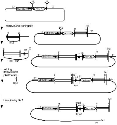
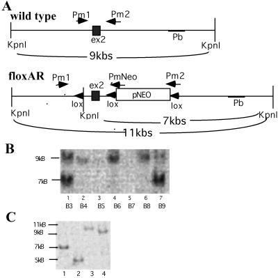
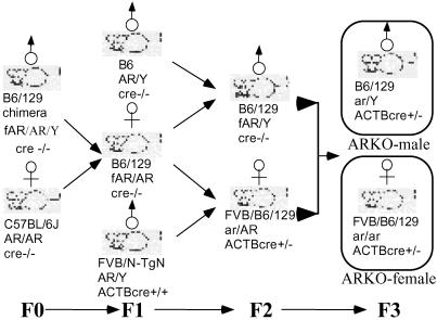
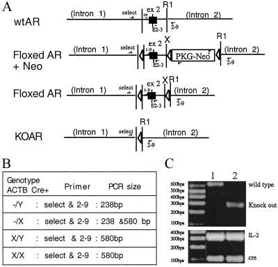
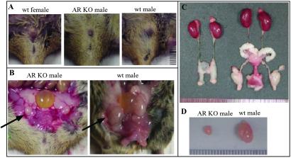
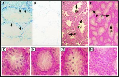
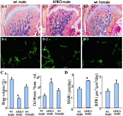
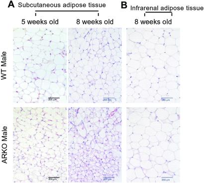
By using a cre-lox conditional knockout strategy, we report here the generation of androgen receptor knockout (ARKO) mice. Phenotype analysis shows that ARKO male mice have a female-like appearance and body weight. Their testes are 80% smaller and serum testosterone concentrations are lower than in wild-type (wt) mice. Spermatogenesis is arrested at pachytene spermatocytes. The number and size of adipocytes are also different between the wt and ARKO mice. Cancellous bone volumes of ARKO male mice are reduced compared with wt littermates. In addition, we found the average number of pups per litter in homologous and heterozygous ARKO female mice is lower than in wt female mice, suggesting potential defects in female fertility and/or ovulation. The cre-lox ARKO mouse provides a much-needed in vivo animal model to study androgen functions in the selective androgen target tissues in female or male mice.


Figures

Figure 1
Figure 1
The construction of the floxed AR fragment. The PKI vector is modified from the pBluescript plasmid. It contains a T7 promoter at the 3′ end, a T3 promoter at the 5′ end, two multiple cloning sites (MCS), two lox sites (▹), a positive Neo selective marker (PKG-Neor), and a negative thymidine kinase selective marker (MCT-TK). For the cloning, the XhoI site at the 5′ end MCS was first destroyed. A 3-kb intron 2 fragment was introduced into the 3′ EcoR1 cloning site (R1), followed by a fragment containing intron 1, exon 2, and a small fragment of intron 2 sequences into 5′ XbaI site (X). A lox sequence plus an artificial KpnI site were finally inserted into the XhoI site shortly 5′ to the beginning of exon 2. The constructed plasmid was linearized by NotI before being electroporated into ES cells.
Figure 2
Figure 2
Screening of the extracted DNA to distinguish wtAR from floxed AR. (A) AR fragment and the flanking region. The restriction fragment of KpnI in wt is 9 kb. Three lox sites and an artificial KpnI restriction site are in the floxed AR fragment. The KpnI restriction resulted in one 7-kb and one 4-kb fragment in floxed AR. By using the 3′ end sequence as the probe (Pb), the Southern blot hybridization displayed a 9-kb fragment from ES cell clones containing wtAR, and a strong 7-kb fragment from specifically recombined ES cells containing floxed AR plus a weak 9-kb fragment from cocultured STO cells. (B) Southern blot screening of ES cells transfected with floxed AR: B3 and B9 clones are recombinated specifically and displayed a strong signal at the 7-kb position from ES cells with floxed AR and a weak signal at the 9-kb position from cocultured STO cells. B4 to B8 are the wt ES clones that displayed signal only at the 9-kb position. (C) Southern blot screening of the floxed AR clone transfected with pCMV plasmid. The pCMV-Cre restricted the sequence between two lox sites and generated four types. Lane 1 is without recombination (7 kb), lane 2 is type I recombination (5 kb), lane 3 is type II (11 kb), and lane 4 is the type III recombination (9 kb).
Figure 3
Figure 3
Generation of ARKO female mice by mating among floxed AR founders and cre founders. The chimera founder is B6/129 mosaic strain. The mating of the founder with the B6 female created female mice heterozygous with floxed AR. The following F2 generation generated floxed AR male mice. The mating between the heterozygous floxed AR female and the homozygous FVB/N-TgN ACTB cre male that carry Cre under β-actin promoter created a heterozygous KO of AR female carrying the Cre. The mating between the floxed AR male and the heterozygous ARKO female carrying the Cre generated the homozygous ARKO Cre+ female mice with a 1:8 ratio.
Figure 4
Figure 4
Genotyping of ARKO mice. We have applied the primers “select” and “2–9” to identify wt and ARKO male mice in our study. (A) Schematic presentation of the DNA construct and primer location in exon 2 area of wt, KO, and floxed AR genes. (B) List of the sizes of PCR product amplified by designed primer pairs. (C) The identification of wt and ARKO mice, by using select and 2–9. We amplified a DNA fragment with 580 bp, which represents wtAR, and with 238 bp, which represents ARKO exon 2. The expression of Cre and internal control IL-2 were confirmed by PCR (Bottom).
Figure 5
Figure 5
Phenotype of 8-week-old male ARKO mice. Six 8-week-old male ARKO mice were killed. The results were always compared among siblings. (A) The external genitalia of male ARKO, wt male, and wt female. (B and C) The internal genitalia of male ARKO and wt male mice. Arrows in B identify the testis. (D) The testes of wt male and ARKO mice.
Figure 6
Figure 6
Immunostaining of AR and histologic analysis of testes from wt and ARKO male mice. Immunostaining of AR in testes from wt (A) and ARKO mice (B). For AR staining, anti-AR antibody and Vectorstain ABC-AP kit were used according to the manufacturer's procedures. Testes from wt show positive signal (A, arrows), and testes from ARKO mice show no positive staining. (×40) The histology of the testis from wt (C) and ARKO (D) shown by hematoxylin and eosin stain. Spermatogenesis in ARKO mice arrested in pachytene spermatocyte (P) stage. (×40) Rs, round spermatids; Es, elongated spermatids; P, pachytene spermatocytes. In further analysis of testes in complete ARKO mice we obtained the following information (E–H): (E) The tubule contains hypoplastic spermatogonia and pachytene spermatocyte. (F) The tubule contains Sertoli cells only and shows fibrillary degeneration in the cytoplasm of Sertoli cells. (G) Some cells in the tubule contain condensed and pyknotic nuclei. They may represent apoptotic bodies. (H) Leydig cells located in interstitial space are hypertrophic.
Figure 7
Figure 7
Bone phenotype in ARKO mice. (A) Hematoxylin and eosin-stained femoral bones of 8-week-old wt and ARKO mice. Note the decreased trabecular bone area in the distal metaphysis of the ARKO mouse compared with wt littermates. (B) Calcein double labeling in 8-week-old wt and ARKO mice. The mean distance between the two yellow labels (white lines) represents the rate of bone mineralization from which the rate of bone formation is calculated. (C) Bone volume and osteoclast numbers data in ARKO and wt mice. Data are from six ARKO male mice and the same number of male and female littermates. (D) Mineral apposition and bone formation rate data in ARKO and wt mice. Data are from three ARKO mice and the same number of wt littermates. *, statistically significant differences compared with control mice (P < 0.05). Error bars represent SD.
Figure 8
Figure 8
Histology of fat tissues in ARKO mice. (A) s.c. adipose tissue of 5- and 8-week-old ARKO and wt littermates were stained with hematoxylin and eosin. (Bars = 200 μm.) (B) Infrarenal adipose tissue of 8-week-old ARKO and wt littermates were stained with hematoxylin and eosin. (Bars = 200 μm.)

Similar articles

Cited by

References

    1. Chang C S, Kokontis J, Liao S T. Science. 1988;240:324–326. - PubMed
    1. Chang C S, Kokontis J, Liao S T. Proc Natl Acad Sci USA. 1988;85:7211–7215. - PMC - PubMed
    1. Lubahn D B, Joseph D R, Sullivan P M, Willard H F, French F, Wilson E M. Science. 1988;240:327–330. - PubMed
    1. Chang C, Saltzman A, Yeh S, Young W, Keller E, Lee H J, Wang C, Mizokami A. Crit Rev Eukaryotic Gene Expression. 1995;5:97–125. - PubMed
    1. Heinlein C A, Chang C. Endocr Rev. 2002;23:175–200. - PubMed

Publication types

MeSH terms

LinkOut - more resources