. 2002 Oct 29;99(22):14298-302.
doi: 10.1073/pnas.162491399. Epub 2002 Oct 7.

Uptake of the anticancer drug cisplatin mediated by the copper transporter Ctr1 in yeast and mammals

Affiliations

Uptake of the anticancer drug cisplatin mediated by the copper transporter Ctr1 in yeast and mammals

Seiko Ishida et al. Proc Natl Acad Sci U S A. .

Abstract

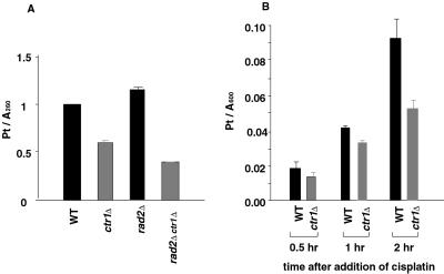
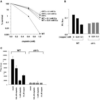
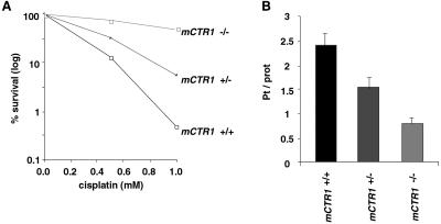
Cisplatin is a chemotherapeutic drug used to treat a variety of cancers. Both intrinsic and acquired resistance to cisplatin, as well as toxicity, limit its effectiveness. Molecular mechanisms that underlie cisplatin resistance are poorly understood. Here we demonstrate that deletion of the yeast CTR1 gene, which encodes a high-affinity copper transporter, results in increased cisplatin resistance and reduced intracellular accumulation of cisplatin. Copper, which causes degradation and internalization of Ctr1 protein (Ctr1p), enhances survival of wild-type yeast cells exposed to cisplatin and reduces cellular accumulation of the drug. Cisplatin also causes degradation and delocalization of Ctr1p and interferes with copper uptake in wild-type yeast cells. Mouse cell lines lacking one or both mouse Ctr1 (mCtr1) alleles exhibit increased cisplatin resistance and decreased cisplatin accumulation in parallel with mCtr1 gene dosage. We propose that cisplatin uptake is mediated by the copper transporter Ctr1p in yeast and mammals. The link between Ctr1p and cisplatin transport may explain some cases of cisplatin resistance in humans and suggests ways of modulating sensitivity and toxicity to this important anticancer drug.


Figures

Fig 1.
Fig 1.
Cisplatin resistance of yeast mutants. Logarithmic-phase cells were exposed for 2 (AC) or 3 (D) h to different concentrations of cisplatin and incubated on YPD plates at 30°C for 2 days to allow formation of colonies. Data are expressed as percentages of colonies formed compared with control cultures not exposed to cisplatin. (A) Cisplatin resistance of mutants defective in Mac1p target genes. Strains are wild-type (YSI1), mac1Δ (YSI9), ctr1Δ (YSI22), fre1Δ (YSI23), fre7Δ (YSI24), cta1Δ (YSI25), and ctt1Δ (YSI26). (B) Cisplatin resistance of mutants defective in intracellular copper trafficking and utilization. Strains are wild-type (YSI1), ctr1Δ (YSI22), atx1Δ (YSI28), ccc2Δ (YSI29), fet3Δ (YSI30), lys7Δ (YSI31), sod1Δ (YSI32), cox17Δ (YSI33), and sco1Δ (YSI34). (C) Cisplatin sensitivity of low-affinity copper transporter mutants, ctr2Δ (YSI35) and fet4Δ (YSI36). (D) Cisplatin sensitivity of high-affinity copper transporter mutants in the BR10 strain background (22). Strains were DTY1 (CTR1 CTR3), SKY52 (ctr1Δ CTR3), SKY44 (CTR1 ctr3Δ), and SKY46 (ctr1Δ ctr3Δ).
Fig 2.
Fig 2.
Cisplatin adduct formation and accumulation in yeast ctr1Δ mutants. (A) Accumulation of cisplatin-DNA adducts in wild-type (WT) and isogenic ctr1Δ mutant strains. Strains were YSI1 (wild-type), YSI22 (ctr1Δ), YSI16 (rad2Δ), and YSI27 (rad2Δ ctr1Δ). Genomic DNA was purified from cells treated with 1 mM cisplatin for 2 h in YPD. Platinum was measured using an atomic absorption spectrometer. Numbers represent absorption by atomized platinum divided by A260 of each DNA sample. (B) Cellular platinum accumulation in wild-type (YSI1) and ctr1Δ mutant (YSI22) strains. The amount of platinum in whole cells was measured using the atomic absorption spectrophotometer after incubating cells with 1 mM cisplatin for the indicated times. Numbers were obtained by dividing the reading from the spectrophotometer by the OD600 of the culture at each time point.
Fig 3.
Fig 3.
A Ctr1p-mediated link between cisplatin and copper transport in yeast. (A) Effect of copper on survival of wild-type (WT) (YSI1) and the ctr1Δ mutant (YSI22) exposed to cisplatin. Cells were treated for 2 h with various concentrations of cisplatin in the presence of 0.01 mM, 0.1 mM, or no CuSO4. Data were analyzed as in Fig. 1. (B) Effect of copper on cellular accumulation of platinum in wild-type and ctr1Δ mutants. Cells were exposed to 1 mM cisplatin in the presence of 0.01 mM, 0.1 mM, or no CuSO4. Data were analyzed as in Fig. 2B. (C) Effect of cisplatin on copper uptake in wild-type and ctr1Δ mutants. Cells were treated with 64Cu (5 μM as CuSO4) in the presence of 100 μM or 1 mM cisplatin, or 10 μM unlabeled CuSO4 for 2 h. Cellular 64Cu level was measured with a γ-counter, and the values were divided by the cell OD600.
Fig 4.
Fig 4.
Effect of cisplatin on yeast Ctr1 protein. (A) Degradation of yeast Ctr1p upon cisplatin treatment. The Ctr1 protein was tagged at its C terminus with an HA epitope by modification of the genomic CTR1 locus (YSI37). Cycloheximide (100 μg/ml) was added to the CTR1-HA cells 30 min before cisplatin treatment to block new protein synthesis. Cells were then exposed to 0.1 mM or no cisplatin, and samples were taken at the indicated times. The level of Ctr1 protein was determined by Western blotting using anti-HA antibodies. The loading of proteins in each lane appeared equal by Coomassie blue staining. (B) Localization of yeast Ctr1p after cisplatin or copper treatment. The Ctr1 protein was tagged at its C terminus with GFP by modification of the genomic CTR1 locus (YSI38). Cells were incubated with 100 μg/ml cycloheximide for 30 min to block new protein synthesis, treated with 1 mM cisplatin for 4 h, and analyzed by fluorescence microscopy.
Fig 5.
Fig 5.
Cisplatin resistance and accumulation by mouse embryonic cell lines lacking mCtr1. Cells were incubated for 2 h with cisplatin. For determining survival (A), cells were stained with trypan blue to detect dead cells; unstained cells were counted using a hemocytometer. Data are expressed as percentages of unstained cells compared with control cultures not exposed to cisplatin. For measuring cisplatin accumulation (B), cells were lysed and, after a centrifugation, the supernatant was used to determine platinum content. The platinum reading was normalized to protein concentration.

Comment in

References

    1. Loehrer P. J. & Einhorn, L. H. (1984) Ann. Intern. Med. 100, 704-713. - PubMed
    1. Giaccone G. (2000) Drugs 59, Suppl. 4, 9-17. - PubMed
    1. Beck D. J. & Brubaker, R. R. (1973) J. Bacteriol. 116, 1247-1252. - PMC - PubMed
    1. Fraval H. N. A., Rawlings, C. J. & Roberts, J. J. (1978) Mutat. Res. 51, 121-132. - PubMed
    1. McA'Nulty M. M. & Lippard, S. J. (1996) Mutat. Res. 362, 75-86. - PubMed

Publication types

MeSH terms