

Case Reports
Granulocytic sarcoma of the temporal bone
Affiliations
- PMID: 12372738
- PMCID: PMC7976787
Item in Clipboard
Case Reports
Granulocytic sarcoma of the temporal bone
AJNR Am J Neuroradiol.
2002 Oct.
Abstract
We report an unusual case of granulocytic sarcoma involving the temporal bone. The occurrence of this tumor usually heralds acute myelogenous leukemia or the onset of the blastic phase of chronic myelogenous leukemia. Recognition of this rare entity is important, because early aggressive chemotherapy can cause regression of the tumor, as in our case, and thus improve patient longevity.
Figures

Initial MR images (600/10 [TR/TE]) obtained at presentation. A, Contrast-enhanced fat-suppressed coronal T1-weighted image shows focal area of enhancement along the anterosuperior surface of the left petrous pyramid (arrow). B, Contrast-enhanced fat-suppressed axial T1-weighted image shows enhancement along the anterosuperior surface of the left petrous pyramid with extension into the geniculate ganglion (long arrow) and the tympanic segment (short arrow) of the left facial nerve. On an adjacent image (not shown), the mass extended into the posterior left cavernous sinus.

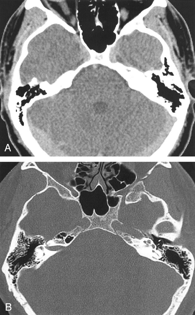
CT scans obtained 2 months later. A, Contrast-enhanced axial CT scan shows a well-defined approximately 2-cm mass (arrow) along the anterosuperior aspect of the left petrous pyramid. Anteriorly, it is in continuity with the cavernous sinus. B, High-spatial-resolution axial CT scan (bone window) shows widening of the left facial nerve canal in the region of the geniculate ganglion (arrow). Opacification of the mastoid air cells by serous fluid is also noted, probably secondary to blockage of the eustachian tube by the petrous apex mass. C, Coronal CT scan, obtained at the level of the cochlea, shows extension of the soft tissue mass into the left middle ear cavity. Erosion of the facial nerve canal at the geniculate ganglion (arrow) is seen.
CT scans obtained after chemotherapy and resolution of symptoms. A, Axial CT scan shows resolution of mass, previously noted along the anterosuperior aspect of the left petrous pyramid. B, Axial CT scan (bone window), obtained at the level of the cochlea, shows no mass within the middle ear cavity and clear mastoid air cells. The anterior portion of the left carotid canal is faintly seen, and this could represent a residual (erosive) change from the previously noted mass.
References
-
- Burns A. Observations of Surgical Anatomy: Head and Neck. Edinburgh: Thomas Royce and Company;1811:364–366
-
- Dock G. Chloroma and its relation to leukemia. Am J Med Sci 1893;106:152–157
-
- Neiman RS, Barcos M, Berard C, et al. Granulocytic sarcoma: a clinicopathologic study of 61 biopsied cases. Cancer 1981;48:1426–1437 - PubMed
-
- Azzarelli B, Roessmann U. Pathogenesis of central nervous system infiltration in acute leukemia. Arch Pathol Lab Med 1977;101:203–205 - PubMed
-
- Cho JS, Kim EE, Ro JH, Pinkel DP, Goepfert H. Mandibular chloroma demonstrated by magnetic resonance imaging. Head Neck 1990;12:507–511 - PubMed
Publication types
MeSH terms
LinkOut - more resources
Full Text Sources