. 2002 Oct;23(9):1500-8.

The craniocervical venous system in relation to cerebral venous drainage

Affiliations

The craniocervical venous system in relation to cerebral venous drainage

Diego San Millán Ruíz et al. AJNR Am J Neuroradiol. 2002 Oct.

Abstract

Background and purpose: Passing from the supine to the upright position favors cerebral venous outflow into vertebral venous systems rather than into the internal jugular veins. We sought to determine venous connections between dural venous sinuses of the posterior cranial fossa and craniocervical vertebral venous systems.

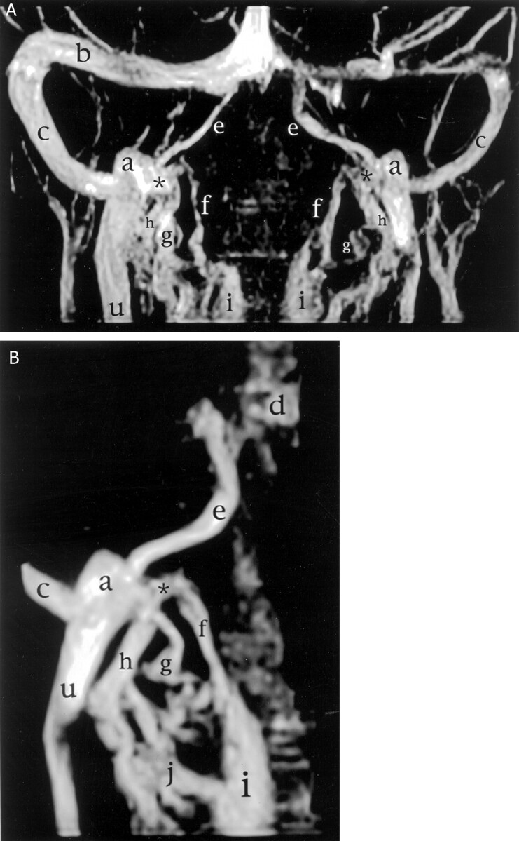
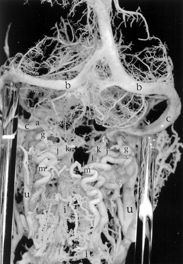
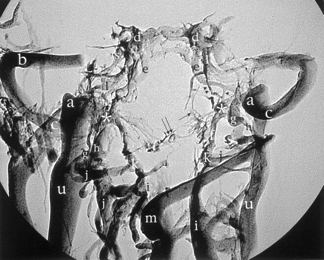
Methods: Corrosion casts of the cranial and cervical venous system were obtained from 12 fresh human cadavers, and anatomic confirmation was made by dissection of three previously injected fresh human specimens. MR venography was performed to provide radiologic correlation.

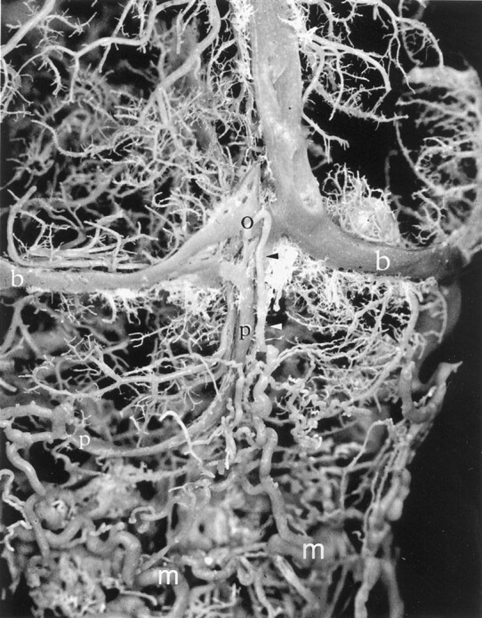
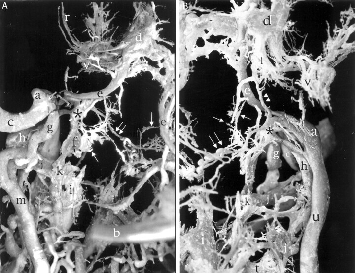
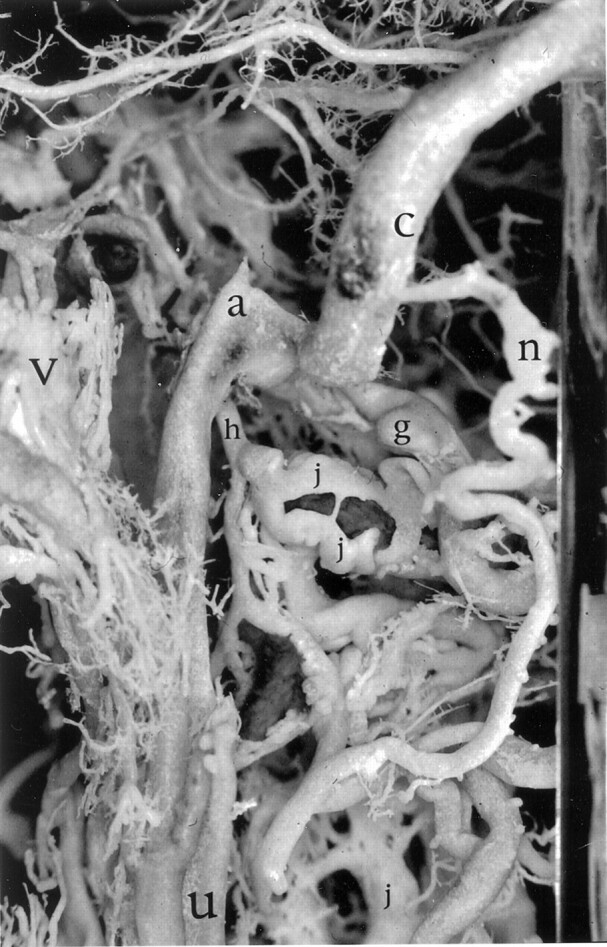
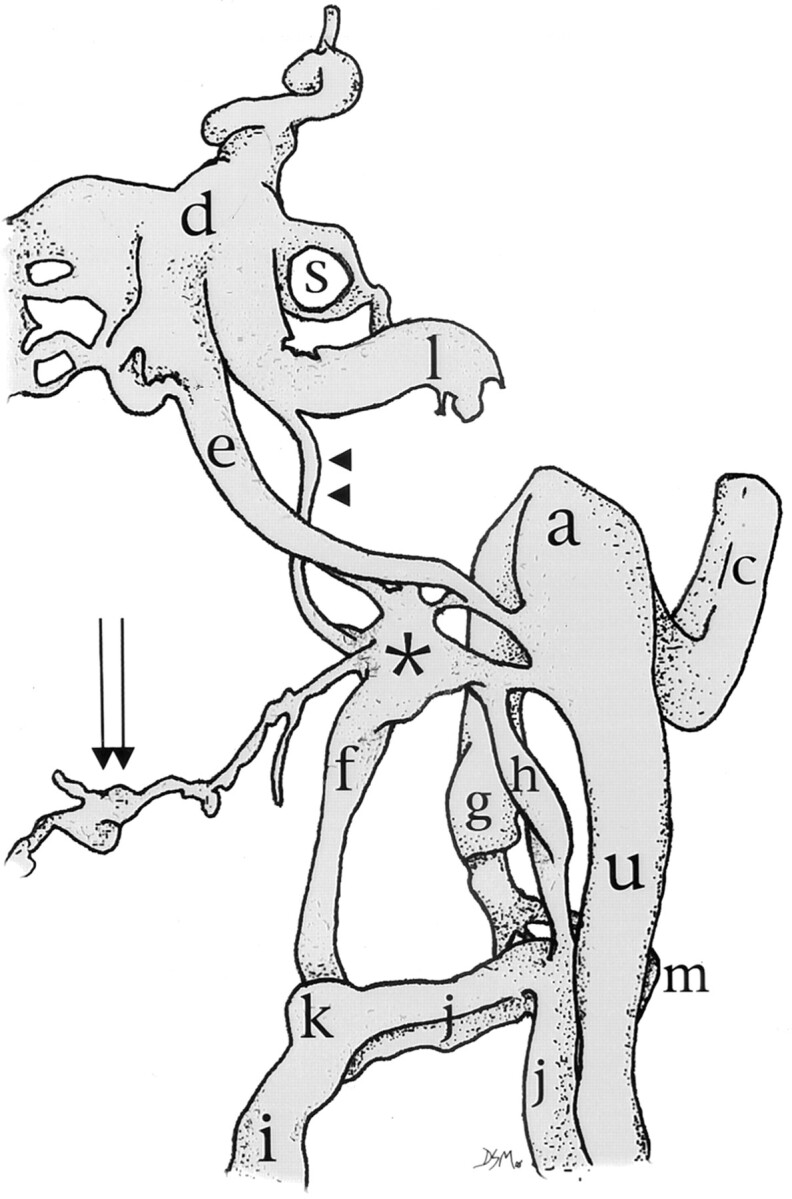
Results: The lateral, posterior, and anterior condylar veins and the mastoid and occipital emissary veins were found to represent the venous connections between the dural venous sinuses of the posterior cranial fossa and the vertebral venous systems. This study revealed the nearly constant presence of the anterior condylar confluent (ACC) located on the external orifice of the canal of the hypoglossal nerve. The ACC offered multiple connections with the dural venous sinuses of the posterior cranial fossa, the internal jugular vein, and the vertebral venous system. All these structures were shown by MR venography.

Conclusion: The lateral, posterior, and anterior condylar veins and the mastoid and occipital emissary veins connect the dural venous sinuses of the posterior cranial fossa with the vertebral venous systems. These connections are clinically relevant, because encephalic drainage occurs preferentially through the vertebral venous system in the upright position. The ACC is a constant anatomic structure that may play an important role in the redirection of cerebral blood in the craniocervical region.


Figures

F<sc>ig</sc> 1.
Fig 1.
Posterior view of a corrosion cast shows an occipital emissary vein (o) draining into the deep cervical veins and a left occipital sinus (p). Black arrowhead and white arrowhead, extracranial connection of the occipital emissary vein; b, transverse sinus; m, deep cervical vein.
F<sc>ig</sc> 2.
Fig 2.
Left jugular bulb region. The ACC (asterisk) and its connections with surrounding veins are shown. The proximal portions of both transverse sinuses and confluens sinuum have been removed for better visualization. Double arrowhead, inferior petrooccipital vein; arrow, basilar plexus; double arrow, branch to prevertebral venous plexus; r, middle meningeal veins; d, cavernous sinus; a, superior jugular bulb; e, inferior petrosal sinus; c, sigmoid sinus; g, posterior condylar vein; h, lateral condylar vein; f, anterior condylar vein; j, vertebral artery venous plexus; k, anastomosis between anterior internal vertebral venous plexus and vertebral artery venous plexus; i, anterior internal vertebral venous plexus; m, deep cervical vein; b, transverse sinus; l, internal carotid artery venous plexus of Rektorzik; s, emissary vein of the foramen ovale; v, pterygoid plexus; t, intervertebral veins, including inter atlanto-occipital vein. A, Posterior view. B, Anterior view.
F<sc>ig</sc> 3.
Fig 3.
MR venograms of the craniocervical region (2D time-of-flight gradient echo images with magnetization transfer; 40/7; field of view, 15 cm; 18; flip angle, 35 degrees; resolution, 220/256; section thickness, 1.5 mm; section overlap, 0.5 mm; matrix, 256 × 256). The ACC (asterisk) and the anterior (f), posterior (g), and lateral (h) condylar veins are shown. b, transverse sinus; c, sigmoid sinus; e, inferior petrosal sinus; a, superior jugular bulb; u, internal jugular vein; i, anterior internal vertebral venous plexus; d, cavernous sinus; j, vertebral artery venous plexus. A, Overview. B, Focused on the right superior jugular bulb.
F<sc>ig</sc> 4.
Fig 4.
Left lateral view of the sigmoid sinus/jugular bulb junction. The mastoid emissary vein (n) and lateral (h) and posterior (g) condylar veins are shown. The plexiform nature of the vertebral artery venous plexus (j), shown here around the horizontal portion of V3, is well seen. c, sigmoid sinus; a, superior jugular bulb; v, pterygoid plexus; u, internal jugular vein.
F<sc>ig</sc> 5.
Fig 5.
Posterior overview of the craniocervical junction in a venous corrosion cast. Note prominent deep cervical veins bilaterally (m). The left posterior condylar vein (g) is clearly seen to drain into both the horizontal portion of the left vertebral artery venous plexus at the C1 level (j) and the left deep cervical vein (m). b, transverse sinus; c, sigmoid sinus; a, superior jugular bulb; f, anterior condylar vein; k, anastomosis between anterior internal vertebral venous plexus and vertebral artery venous plexus; i, anterior internal vertebral venous plexus; u, internal jugular vein.
F<sc>ig</sc> 6.
Fig 6.
Right lateral view of the craniocervical junction in a venous corrosion cast. Note the presence of a prominent mastoid emissary vein (n) connecting to a deep cervical vein (m). The carotid artery venous plexus is clearly visible (l). b, transverse sinus; d, cavernous sinus; c, sigmoid sinus; a, superior jugular bulb; v, pterygoid plexus; g, posterior condylar vein; h, lateral condylar vein; u, internal jugular vein.
F<sc>ig</sc> 7.
Fig 7.
Standard radiographic picture of the corrosion cast from Figure 2 in the anteroposterior projection shows the ACC (asterisk) and its relation with surrounding veins. The proximal portions of the transverse sinus (b), confluens sinuum, and straight and superior longitudinal sinuses have been removed for better visualization. Arrowhead, branch between the inferior petrosal sinus and the ACC; double arrowhead, inferior petrooccipital vein; arrow, basilar plexus; double arrow, branch to prevertebral venous plexus; d, cavernous sinus; e, inferior petrosal sinus; l, internal carotid artery venous plexus of Rektorzik; a, superior jugular bulb; g, posterior condylar vein; c, sigmoid sinus; h, lateral condylar vein; k, anastomosis between anterior internal vertebral venous plexus and vertebral artery venous plexus; j, vertebral artery venous plexus; u, internal jugular vein; i, anterior internal vertebral venous plexus; m, deep cervical vein.
F<sc>ig</sc> 8.
Fig 8.
Schematic representation of anterior view of the ACC and its connections. Note the six main contributions, from the anterior condylar vein (f), the lateral condylar vein (h), the internal jugular vein (u), the inferior petrosal sinus (e), the venous plexus of Rektorzik (double arrowhead), and the prevertebral venous plexus (double arrow). d, cavernous sinus; s, emissary vein of the foramen ovale; a, superior jugular bulb; c, sigmoid sinus; g, posterior condylar vein; k, anastomosis between anterior internal vertebral venous plexus and vertebral artery venous plexus; j, vertebral artery venous plexus; m, deep cervical vein; i, anterior internal vertebral venous plexus.

References

    1. Hacker H. Superficial supratentorial veins and dural sinuses. In: Potts DG, Newton TH, eds. Radiology of the Skull and Brain. vol 2, book 3. St Louis: The CV Mosby Company;1974. :1851–1877
    1. Breschet G. Recherches Anatomiques, Physiologiques et Pathologiques sur le Système Veineux et Spécialement sur les Canaux Veineux des Os [in French]. Paris: Villeret et Rouen;1829:1–42
    1. Clemens H. 1. Beitrag zur Histologie des Plexus venosi vertebrales interni. Zeitschrift für mikr.-anat Forshung 1961;67:183–189
    1. Batson O. The vertebral vein system. AJR Am J Roentgenol 1957;78:195–212 - PubMed
    1. Groen RJ, Groenewegen HJ, van Alphen HA, Hoogland PV. Morphology of the human internal vertebral venous plexus: a cadaver study after intravenous Araldite CY 221 injection. Anat Rec 1997;249:285–294 - PubMed