

Reproducibility of primary motor cortex somatotopy under controlled conditions
- PMID: 12372742
- PMCID: PMC7976799
Reproducibility of primary motor cortex somatotopy under controlled conditions
Abstract
Background and purpose: The somatotopic organization of the contralateral primary motor cortex (M1) and its intra- and intersubject reproducibility has been the subject of many investigations and controversies. A potential explanation for a least some of the conflicting results could be the lack of movement control in the studies performed. The purpose of this study was to investigate these issues under controlled experimental conditions.
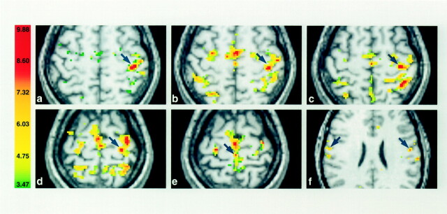
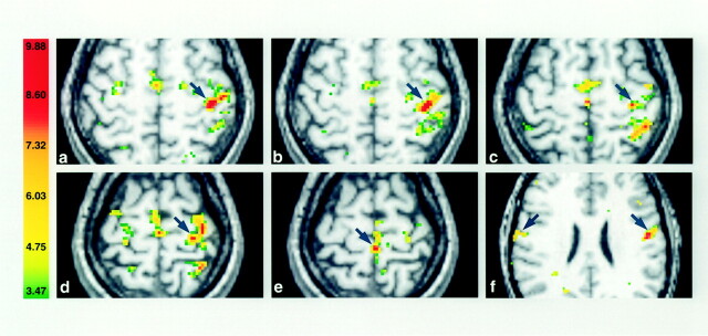
Methods: Functional MR imaging was performed in 12 healthy volunteers performing hand, finger, wrist, elbow, foot, and tongue movements. Two experimental sessions were separated by 2 weeks. Controlled conditions were achieved by means of a custom-designed arm and hand manipulandum providing standardization of the movements within and across subjects.
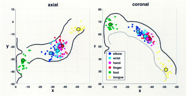
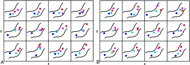
Results: The experiments revealed a clear large-scale somatotopy of the contralateral M1 with distinct subregions controlling the foot, arm, and tongue. Despite considerable overlap of the volumes, geometric centers of gravity (COGs) showed statistically significant differences in coordinates between the elbow, wrist, fingers, and hand. COGs showed a high degree of intra- and interindividual reproducibility, particularly for the upper limb movements, in contrast to the activation volumes that proved to be unreliable parameters, despite the controlled conditions.
Conclusion: These findings support the existence of a gross-scale somatotopic organization yet also demonstrate a clear, fine-scale somatotopy of the within-arm representations. Furthermore, they reveal high reproducibility of the COGs when standardized conditions are applied. This observation highlights the need for movement control to allow for intra- and intersubject comparison.
Figures
References
-
- Hughlings Jackson J Convulsive spasms of the right hand and arm preceding epileptic seizures. Med Times Gaz 1863;1:589
-
- Foerster O. Motorische Felder und Bahnen. In: Bumke H, Foerster O, eds. Handbuch der Neurologie IV. Berlin: Springer-Verlag;1936;49–56
-
- Penfield W, Boldrey E. Somatic motor and sensory representation in the cerebral cortex of man as studies by electrical stimulation. Brain 1937;60:389–443
-
- Hepp-Reymond MC. Functional organization of motor cortex and its participation in voluntary movements. In: Setklis HD, Erwin J, eds. Comparative Primate Biology Volume 4: Neurosciences, New York: Alan R. Liss, Inc;1988. :501–624
-
- Lemon R. Mapping the output functions of the motor cortex. In: Edelman G, Gall W, Cowan W, eds. Signal and Sense New York: Wiley-Liss;1990. :315–355
MeSH terms
LinkOut - more resources
Full Text Sources
Medical