

Case Reports
Rapid enlargement of a posterior communicating artery aneurysm after Guglielmi detachable coil treatment of ipsilateral carotid artery aneurysms
Affiliations
- PMID: 12372751
- PMCID: PMC7976785
Item in Clipboard
Case Reports
Rapid enlargement of a posterior communicating artery aneurysm after Guglielmi detachable coil treatment of ipsilateral carotid artery aneurysms
AJNR Am J Neuroradiol.
2002 Oct.
Abstract
This case illustrates rapid aneurysm enlargement, presumably due to altered hemodynamics resulting from endovascular treatment of aneurysms on the same artery. We postulate that increased hemodynamic force directed to the inflow zone of the posterior communicating artery aneurysm was caused by the treatment of the two ophthalmic artery aneurysms. Originally, many of the flow vectors may have been directed into the larger ophthalmic segment aneurysm, located on the outside of the curve of the internal carotid artery. After treatment, flow may have been directed more smoothly around the carotid siphon and into the posterior communicating artery aneurysm.
Figures
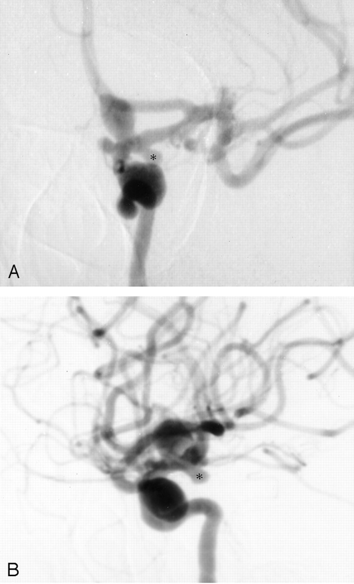
Angiograms from the case of a 45-year-old woman with chronic headaches who presented after experiencing a fall. Five aneurysms were present on the left internal carotid artery. A, Anteroposterior view angiogram of the left internal carotid artery shows multiple aneurysms. These include a large cavernous carotid aneurysm, a small ophthalmic segment aneurysm, a large ophthalmic segment aneurysm, and a posterior communicating artery aneurysm (asterisk). A tiny anterior choroidal aneurysm is also present. B, Lateral view angiogram of the left internal carotid artery. Astserisk indicates posterior communicating artery aneurysm.
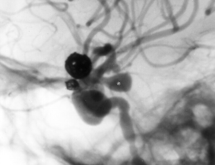
Four months after GDC treatment of the two ophthalmic segment aneurysms, lateral view angiogram of the left carotid artery shows marked interval enlargement of the posterior communicating artery aneurysm (asterisk, compare with Fig 1B). The treated ophthalmic aneurysms remain completely obliterated.
References
-
- Ferguson GG. Physical factors in the initiation, growth, and rupture of human intracranial saccular aneurysms. J Neurosurg 1972;37:666–677 - PubMed
-
- Misra BK, Whittle IR, Steers AJ, Sellar RJ. De novo saccular aneurysms. Neurosurgery 1988;23:10–15 - PubMed
-
- Stehbens WE. Etiology of intracranial berry aneurysms. J Neurosurg 1989;70:823–831 - PubMed
Publication types
MeSH terms
LinkOut - more resources
Full Text Sources
Medical