. 2002 Oct 7;87(8):859-67.
doi: 10.1038/sj.bjc.6600545.

Binding of TGF-beta1 latency-associated peptide (LAP) to alpha(v)beta6 integrin modulates behaviour of squamous carcinoma cells

Affiliations

Binding of TGF-beta1 latency-associated peptide (LAP) to alpha(v)beta6 integrin modulates behaviour of squamous carcinoma cells

G J Thomas et al. Br J Cancer. .

Abstract

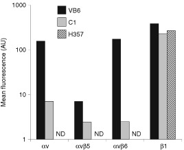
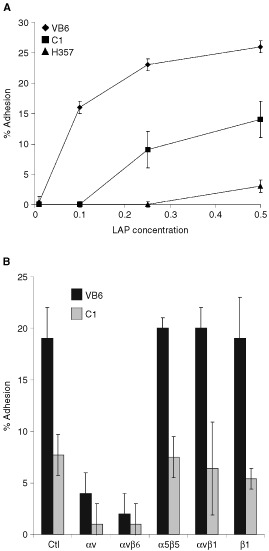
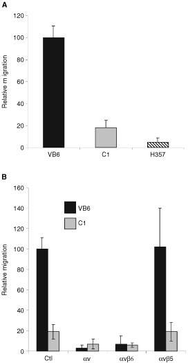
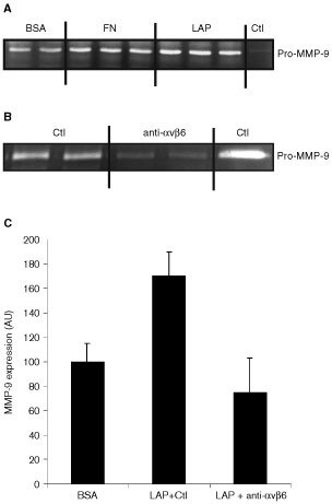
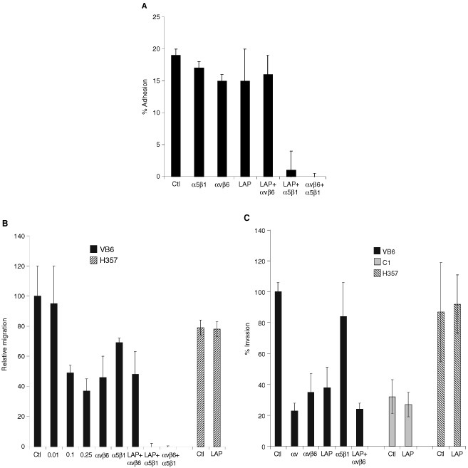
The integrin alpha(v)beta6 is not detectable on normal keratinocytes in vivo but expression is increased significantly in oral squamous cell carcinoma where this heterodimer has been shown to play a role in cell migration, invasion and protease expression. Although regarded initially as a fibronectin receptor, alpha(v)beta6 may bind to arginine-glycine-aspartic acid sequences in other matrix molecules including tenascin and vitronectin. Interestingly, alpha(v)beta6 has also been shown to have high affinity for the TGF-beta1 latency associated peptide and to participate in the activation of the TGF-beta1 latent complex. Since TGF-beta1 is present in squamous carcinomas, it is possible that latency associated peptide may modulate malignant keratinocyte behaviour independently from the classical TGF-beta signalling pathways through its interaction with integrins. We show here that when latency associated peptide is immobilised onto a surface, it acts as an alpha(v)beta6-specific ligand for oral squamous carcinoma cells promoting adhesion and haptotactic migration in addition to alpha(v)beta6-dependent increase in pro-MMP-9 expression. In contrast, even very low concentrations of soluble latency associated peptide (0.1 microg ml(-1)) inhibited alpha(v)beta6-dependent adhesion, migration and invasion. Thus alpha(v)beta6-dependent processes of oral squamous cell carcinoma, is likely to be modulated, not only by the local concentration of latency associated peptide in the stroma, but also whether it is immobilised in the matrix or released as a soluble protein.


Figures

Figure 1
Figure 1
Flow cytometric analysis of integrin expression by cell lines. The geometric mean fluorescence (arbitrary units, log scale) as measured by flow cytometry of cells labelled with anti-integrin antibodies is shown. Negative control had secondary antibody only and has been subtracted from the results. Figure shows a representative experiment (ND=not detected). Flow cytometry confirmed high αvβ6 expression by VB6 cells and that C1 null transfectant cells express low levels of endogenous αvβ6. H357 cells are αv- negative. The cell lines express similar levels of β1 integrins.
Figure 2
Figure 2
Cell adhesion to LAP is αvβ6-dependent. Chromium [51Cr]- labelled cells (1.5×10⁴) were added to LAP-coated 96-well plates containing an irrelevant control antibody (W632 anti-MHC class 1) or test antibodies against αv (L230), αvβ5 (P1F6), αvβ6 (10D5), β1 (P4C10) and α5β1 (P1D6). Background binding to BSA has been subtracted from the results. Figures show representative experiments performed in quadruplicate. Error bars represent standard deviation. (A) VB6 cells show increased adherence LAP. β6 transfected cells (VB6), control cells (C1) and αv-negative cells (H357) were plated onto varying concentrations of LAP. VB6 cells showed significantly increased adhesion compared to C1 cells (which express low levels of endogenous αvβ6). H357 cells did not adhere. (B) Adhesion to LAP is αvβ6-dependent. VB6 and C1 adhesion to LAP was inhibited by anti-αvβ6 antibody or anti-αv antibody. Antibodies against αvβ5, α5β1 or β1 produced no effect. These data suggest that adhesion of VB6 and C1 cells is modulated solely through αvβ6.
Figure 3
Figure 3
Cell migration towards LAP is αvβ6-dependent. Cells were allowed to migrate towards LAP in haptotactic migration assays. To assess integrin specificity of migration, integrin-blocking antibodies against αv (L230), αvβ5 (P1F6), αvβ6 (10D5), or a control antibody (W632) were added to VB6, C1 and H357 cells prior to plating into wells. Following 8 h incubation, the cells in the lower chamber (including those attached to the undersurface of the membrane) were trypsinised and counted on a Casy 1 counter (Sharfe System GmbH, Germany). Results for the cell lines are expressed relative to VB6 migration following incubation with an irrelevant control antibody (=100). Figures show representative experiments performed in quadruplicate. Error bars represent standard deviation. (A) VB6 cells show increased migration towards LAP. Comparison of migration of VB6, C1 and H357 cells towards LAP. Migration is higher in the αvβ6 expressing VB6 cells. αv-negative H357 cells did not migrate significantly. (B) Migration towards LAP is αvβ6-dependent. Migration of VB6 and C1 cells was inhibited completely by antibody inhibition of αvβ6 or αv. Antibodies inhibiting αvβ5 produced no effect. These data suggest that migration of VB6 and C1 cells is modulated solely through αvβ6.
Figure 4
Figure 4
Binding of αvβ6 to LAP induces upregulation of MMP-9. Zymography for MMP-9. Cells were grown for 24 h in additive-free medium before supernatant sampling and cell counting. Samples containing equal volume per cell number were run on each gel with MMP-9 control. The intensity of the bands was measured by densitometric analysis and comparisons made within each gel to determine relative changes in MMP activity. (A) Zymogram showing upregulation of MMP-9 expression by VB6 cells when plated on fibronectin (FN) and LAP relative to BSA-coated plastic. Control is pro-MMP-9. (B) Zymogram showing the inhibition of MMP-9 expression by VB6 cells plated onto LAP following blockade with anti-αvβ6 antibody 10D5 relative to an irrelevant control antibody (Ctl; W632 (anti-MHC class 1)). (C) Densitometric analysis of zymograms from multiple separate experiments (n=4) showing MMP-9 expression by VB6 cells on BSA, LAP following incubation with an irrelevant control antibody (Ctl; W632 (anti-MHC class 1)) and LAP following blockade with anti-αvβ6 antibody (10D5). Results are expressed relative to MMP-9 expression by VB6 cells on BSA-coated plastic (=100). Error bars represent standard deviation. These data suggest that αvβ6-LAP interaction modulates MMP-9 expression in VB6 cells.
Figure 5
Figure 5
Soluble LAP inhibits αvβ6 interaction with fibronectin. (A) LAP inhibits VB6 adhesion to fibronectin. Chromium [51Cr]-labelled VB6 cells were added to fibronectin-coated 96-well plates containing an irrelevant control antibody (W632) or test antibodies against αvβ6 (10D5), α5β1 (P1D6) and LAP (0.25 μg ml−1) or combinations thereof. Figure shows a representative experiment performed in quadruplicate. Results are expressed relative to binding in the presence of a control antibody (=100). Background binding to BSA has been subtracted from the results. Error bars represent standard deviation. VB6 cells adhere to fibronectin through αvβ6 and α5β1 and a combination of antibodies against both integrins is required to block adhesion completely (Thomas et al, 2001b). Treating the cells solely with LAP, or antibodies against αvβ6 or α5β1 did not inhibit adhesion significantly (neither did a combination of LAP and anti-αvβ6). However, combinations of anti-α5β1 and LAP or anti-αvβ6 and anti-α5β1 inhibited adhesion completely. These data confirm that soluble LAP produces a similar effect to anti-αvβ6 antibody and suggests that LAP may alter keratinocyte binding to fibronectin through its interaction with the αvβ6 integrin. (B) LAP inhibits VB6 migration to fibronectin. To determine the effect of LAP on cell migration, the cells were treated with varying concentrations of LAP peptide prior to plating and then allowed to migrate for 8 h before counting. A representative experiment performed in quadruplicate is shown. Results are expressed relative to migration in the presence of a control antibody (=100). Error bars represent standard deviation. Haptotactic migration of VB6 cells towards fibronectin is modulated through αvβ6 and α5β1 integrins and maximal inhibition of migration is produced by blocking both integrins (Thomas et al, 2001b). Migration of VB6 cells towards fibronectin was inhibited by a concentration of LAP as low as 0.1 μg ml−1 (51% inhibition). At higher concentrations a further degree of inhibition was seen (0.25 μg ml−1=63% inhibition). This level of inhibition was similar to that produced by anti-αvβ6 antibody (54% inhibition) and did not differ significantly from a combination of LAP and anti-αvβ6 antibody (52% inhibition). Complete inhibition of migration was produced using a combination of soluble LAP with anti-α5β1 antibody (or a combination of anti-αvβ6 with anti-α5β1 antibody). Soluble LAP had no effect of the migration of H357 αv-negative cells. These data confirm that soluble LAP produces a similar effect to anti-αvβ6 antibody and suggest that LAP may inhibit αvβ6-dependent keratinocyte migration towards fibronectin. (C) LAP inhibits αvβ6-dependent invasion through Matrigel. Cell invasion assays were performed over 72 h using Matrigel coated polycarbonate filters. To assess the effect of soluble LAP invasion, cells were treated with LAP (0.5 μg ml−1) for 30 min at 4°C prior to plating. Following incubation the cells in the lower chamber (including those attached to the undersurface of the membrane) were trypsinised and counted on a Casy 1 counter (Sharfe System GmbH, Germany). Figure shows a representative experiment performed in quadruplicate. Error bars represent standard deviation. Pre-incubation of VB6 cells with soluble LAP inhibited invasion (62% inhibition), producing a similar level of inhibition to anti-αvβ6 antibody (58% inhibition). Soluble LAP had no effect on invasion of H357 αv-negative cells or C1 control cells which express low levels of endogenous αvβ6. These data suggest that soluble LAP may inhibit αvβ6-dependent cell invasion.

Similar articles

Cited by

References

    1. AhmedNPansinoFClydeRMurthiPQuinnMARiceGEAgrezMVMokSBakerMS2002Overexpression of alphavbeta6 integrin in serous epithelial ovarian cancer regulates extracellular matrix degradation via the plasminogen activation cascade Carcinogenesis 23237244 - PubMed
    1. AkhurstRJBalmainA1999Genetic events and the role of TGF beta in epithelial tumour progression J Pathol 1878290 - PubMed
    1. AkhurstRJDerynckR2001TGF-beta signaling in cancer–a double-edged sword Trends Cell Biol 11S44S51 - PubMed
    1. AnnesJPRifkinDBMungerJS2002The integrin αvβ6 binds and activates latent TGFβ3 FEBS 5116568 - PubMed
    1. ArihiroKKanekoMFujiiSInaiKYokosakiY2000Significance of alpha 9 beta 1 and alpha v beta 6 integrin expression in breast carcinoma Breast Cancer 71926 - PubMed

MeSH terms