. 2002 Oct 7;87(8):909-17.
doi: 10.1038/sj.bjc.6600541.

Apoptotic mechanisms in T47D and MCF-7 human breast cancer cells

Affiliations

Apoptotic mechanisms in T47D and MCF-7 human breast cancer cells

L M Mooney et al. Br J Cancer. .

Abstract

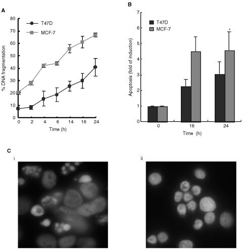
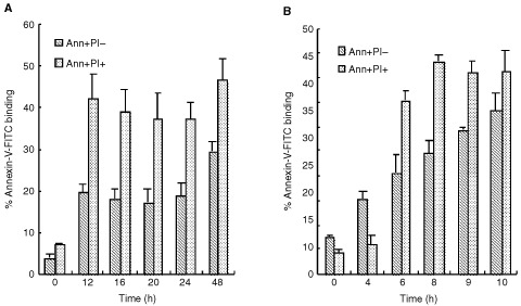
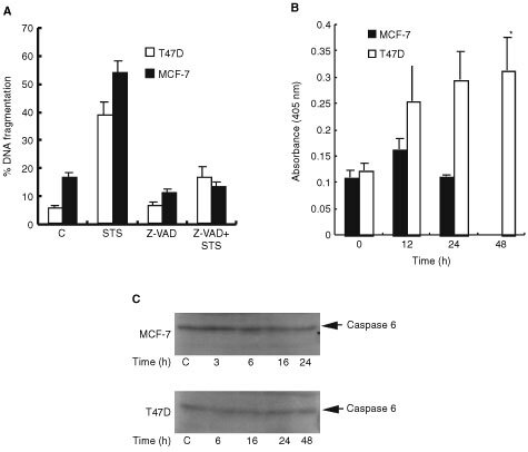
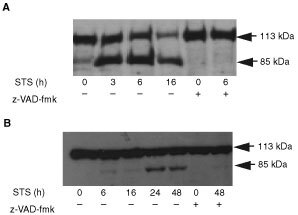
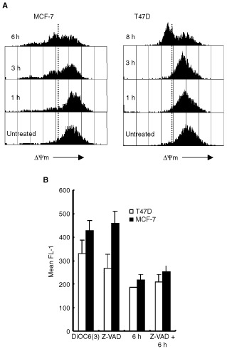
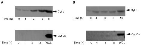
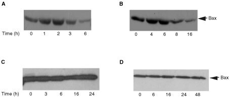
To investigate the mechanisms underlying apoptosis in breast cancer cells, staurosporine was used as an apoptotic stimulus in the human breast cancer cell lines MCF-7 and T47D. Staurosporine induced dose and time dependent increases in DNA fragmentation which was abrogated by z-VAD-fmk. MCF-7 cells did not express caspase-3, suggesting that DNA fragmentation occurred in the absence of caspase-3 and that other caspases may be involved. Staurosporine induced DEVDase activity in T47D cells suggesting the involvement of caspase-3 and/or caspase-7, yet there was no DEVDase activity in MCF-7 cells, probably ruling out the involvement caspase-7. However, staurosporine induced the cleavage of pro-caspase-6 in MCF-7 cells, but not in T47D cells. Caspase dependent PARP cleavage was detected in MCF-7 cells at 3 h, whereas only partial PARP cleavage was detected in T47D cells and then only after 24 h. Moreover, staurosporine led to cytochrome c release at 2 h in MCF-7 cells and 6 h in T47D cells. In addition, a time dependent and caspase-independent reduction of the mitochondrial transmembrane potential was observed; which appeared to occur after the release of cytochrome c. Translocation of Bax from the cytosol to mitochondria was observed in both cell types, and this preceded cytochrome c release in both T47D and MCF-7 cells. Apoptotic events in both cell types differ temporally, involving activation of different caspases and mitochondrial changes.


Figures

Figure 1
Figure 1
Staurosporine induced apoptosis in T47D and MCF-7 human breast cancer cells. (A) Cells were treated with 1 μM STS for the times indicated and apoptosis was determined by DNA fragmentation (ISNT) on fixed and permeabilised cells. Data are presented as per cent DNA fragmentation: error bars represent the mean±s.e.m. of three independent experiments. (B) Cells were treated with 1 μM STS for the times indicated, lysates were analysed by ELISA according to the manufacturer's instructions. Fold induction in apoptosis is expressed as amount of cytoplasm DNA-histone complexes in treated cells compared to untreated controls. Error bars represent the mean±s.e.m. of three independent experiments where * indicates statistically significant increase in apoptosis compared to untreated control (0 h), (ANOVA: F=5.44, P<0.05, d,f 2,9. Tukey multiple comparison test, P<0.05). (C) Apoptotic nuclear morphology. (i) T47D cells (ii) MCF-7 cells. Cells were treated with 1 μM STS for 16 h, then washed in PBS, fixed, permeabilised and stained with 1 μg ml−1 DAPI. Cells were then analysed by fluorescent microscopy.
Figure 2
Figure 2
Staurosporine induced a time dependent increase in annexin-V binding in (A) T47D and (B) MCF-7 cells. Both T47D and MCF-7 cells were treated with 1 μM STS for the times indicated. Cells were washed in PBS and stained with annexin-V-FITC and PI and analysed by Flow Cytometry. Cells staining annexin-V-FITC+/PI- were considered apoptotic; cells staining annexin-V-FITC+/PI+ were classified as late apoptotic/necrotic. Data are presented as per cent of cells in each classification. Error bars represent the mean±s.e.m. of three independent experiments.
Figure 3
Figure 3
Staurosporine induced apoptosis activation of the caspase cascade in both MCF-7 and T47D cells. (A) Cells were pre-incubated with 100 μM z-VAD-fmk (Z-VAD) for 1 h prior to the addition of 1 μM STS for 16 h. Cells were fixed, permeabilised and DNA fragmentation detected by ISNT. Error bars represent the mean±s.e.m. of three independent experiments. (B) Cells were treated for different time periods with 1 μM STS. Cell lysates were prepared and incubated with Ac-DEVD-pNA (caspase-3/7). The reaction products were measured at 405 nm after a 4 h incubation. Error bars represent the mean±s.e.m. of four independent experiments where * indicates statistically significant increase in DEVDase activity compared to untreated control (0 h), (ANOVA: F=3.38, P<0.05, d,f 3,12. Tukey multipe comparison test, P<0.05). (C) Full length caspase-6 protein is cleaved in MCF-7 but not in T47D cells after treatment. Cells were treated with 1 μM STS for indicated times, lysates prepared and 50 μg protein subjected to SDS–PAGE followed by Western transfer and probed with caspase-6 antibody. C indicates untreated cells. The results are representative of three independent experiments.
Figure 4
Figure 4
Staurosporine induced PARP cleavage in MCF-7 and T47D cells. Both (A) MCF-7 and (B) T47D cells were either treated with 1 μM STS for the times indicated or pre-incubated with 100 μM z-VAD-fmk for 1 h prior to STS treatments. Cell lysates (40 μg protein) were prepared and samples subjected to 8% SDS–PAGE followed by Western transfer and probed with PARP antibody (full length protein is 113 kDa, and cleaved fragment is 85 kDa). The results are representative of three independent experiments.
Figure 5
Figure 5
Alterations of the mitochondrial transmembrane potential (ΔΨm) in MCF-7 and T47D cells in response to STS. (A) Cells were treated with 1 μM STS for the times indicated. Cells were then stained with 40 nM DiOC6(3). Representative histoplots are shown for each time point. The dotted line separates cells with high and low fluorescence. (B) Caspase independent ΔΨm collapse. Cells were incubated with 100 μM z-VAD-fmk (Z-VAD) prior to 1 μM STS for 6 h. Mean fluorescence (FL-1) representing ΔΨm was measured by Flow Cytometry. Error bars represent the mean±s.e.m. of four independent experiments.
Figure 6
Figure 6
Staurosporine induced cytochrome c release into the cytosol of (A) MCF-7 and (B) T47D cells. After treatment with 1 μM STS for the times indicated cytosolic extracts were prepared and 30 μg protein subjected to 14% SDS–PAGE followed by Western blot analysis with anti-cytochrome c antibody (Cyt c) and cytochrome c oxidase subunit II (Cyt Ox). Whole cell lysate (WCL) was used as a positive control for the cytochrome c oxidase. Results are representative of four independent experiments.
Figure 7
Figure 7
Staurosporine induced Bax redistribution from the cytosol to the mitochondria in breast cancer cells (A) MCF-7 (B) T47D. After incubation with 1 μM STS for the indicated times cytosolic extracts were prepared and subjected to 14% SDS–PAGE followed by Western blot analysis and probed with anti-Bax or anti cytochrome c oxidase subunit II antibody. Also shown are the Bax levels in the whole cell lysate of (C) MCF-7 and (D) T47D cells respectively. Results are representative of three independent experiments.

References

    1. BlancCDeverauxQLKrajewskiSJanickeRUPorterAGReedJCJaggiRMartiA2000Caspase 3 is essential for procaspase 9 processing and cisplatin induced apoptosis of MCF-7 breast cancer cells Cancer Res 6043864390 - PubMed
    1. Bossy-WetzelENewmeyerDDGreenDR1998Mitochondrial cytochrome c release in apoptosis occurs upstream of DEVD-specific caspase activation and independently of mitochondrial transmembrane depolarization EMBO J 173749 - PMC - PubMed
    1. BurowMEWeldonCBTangYNavarGLKrajewskiSReedJCHammondTGClejanSBeckmanBS1998Differences in susceptibility to tumor necrosis factor α-induced apoptosis among MCF-7 breast cancer cell variants Cancer Res 5849404946 - PubMed
    1. ChittendenTHarringtonEAO'ConnorRFlemingtonCLutzRZEvanGIGuildBC1995Induction of apoptosis by the Bcl-2 homologue Bak Nature 374733736 - PubMed
    1. DecaudinDGeleySHirschTCastedoMMarchettiPMachoAKoflerKKroemerG1997Bcl-2 and Bcl-xl antagonize the mitochondrial dysfunction preceding nuclear apoptosis induced by chemotherapeutic agents Cancer Res 576267 - PubMed

Publication types

MeSH terms