Comparative Study
. 2002 Oct 15;21(20):5323-30.
doi: 10.1093/emboj/cdf537.

Ionic regulation of MscK, a mechanosensitive channel from Escherichia coli

Affiliations
Comparative Study

Ionic regulation of MscK, a mechanosensitive channel from Escherichia coli

Yuezhou Li et al. EMBO J. .

Abstract

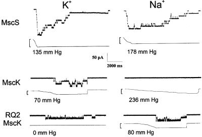
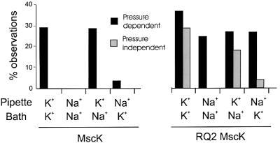
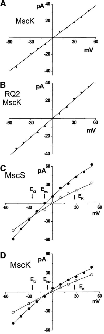
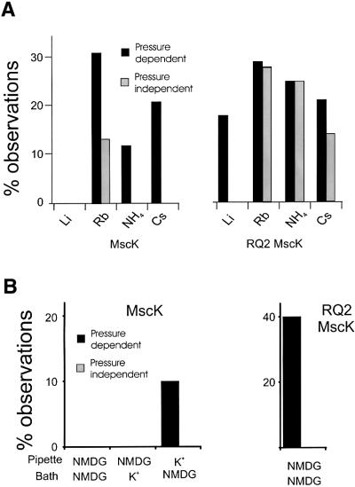
Three gene products that form independent mechanosensitive channel activities have been identified in Escherichia coli. Two of these, MscL and MscS, play a vital role in allowing the cell to survive acute hypotonic stress. Much less is known of the third protein, MscK (KefA). Here, we characterize the MscK channel activity and compare it with the activity of its structural and functional homologue, MscS. While both show a slight anionic preference, MscK appears to be more sensitive to membrane tension. In addition, MscK, but not MscS activity appears to be regulated by external ionic environment, requiring not only membrane tension but also high concentrations of external K(+), NH(4)(+), Rb(+) or Cs(+) to gate; no activity is observed with Na(+), Li(+) or N-methyl-D-glucamine (NMDG). An MscK gain-of-function mutant gates spontaneously in the presence of K(+) or similar ions, and will gate in the presence of Na(+), Li(+) and NMDG, but only when stimulated by membrane tension. Increased sensitivity and the highly regulated nature of MscK suggest a more specialized physiological role than other bacterial mechanosensitive channels.


Figures

None
Fig. 1. Typical traces of MscS (top) and wild-type MscK (middle) and the RQ2 MscK in ionic conditions in which K+ (left) or Na+ (right) is the primary permeant cation. Recordings were generated as described in Materials and methods at –20 mV; channel openings are downward. The bottom trace of each set depicts the pressure in mm Hg; the maximum pressures achieved across the membrane patch are indicated.
None
Fig. 2. The activities of wild-type MscK (left) and the RQ2 MscK (right) are dependent upon the ionic composition of the pipette (periplasmic) solution. The major permeant cation within the pipette or bath is as labelled. The histogram shows the percentage of MscL-containing traces where activities were observed in a spontaneous, pressure- independent (grey) or pressure-dependent (black) manner for each condition. Each percentage is derived from ≥50 MscL-containing traces.
None
Fig. 3. Ionic preferences of wild-type MscK, RQ2 MscK and MscS. (A) The wild-type MscK does not discriminate between K+ or Na+ cations as indicated by the current–voltage relationship of single channel activities when the major permeant cation is K+ (200 mM) within the pipette and Na+ (200 mM) within the bath. (B) In an experiment analogous to that shown in (A), the current–voltage relationship of the RQ2 MscK demonstrates that it also does not discriminate between K+ or Na+ cations. (C) MscS shows an anionic preference as indicated by the current–voltage relationship. Single-channel activities are plotted under symmetrical conditions in which both the pipette and bath contained 200 mM KCl (open circles), then subsequent replacement of the bath with the same buffer containing 600 mM KCl (closed circles) (D) In an analogous experiment to that shown in (C), MscK shows a very slight but measurable anionic preference.
None
Fig. 4. The influence of different ions on the activities of wild-type and RQ2 MscK. In each of the experiments shown, the buffers were as described in Materials and methods and the primary permeant ion is indicated; all were at 200 mM final concentration. (A) The ionic influence on wild-type MscK (left) and the RQ2 MscK (right) activities depends upon the ionic radius of the inorganic cation. The major permeant cation is as labelled. The histogram shows the percentage of MscL-containing traces where activities were observed in a spontaneous, pressure-independent (grey) or pressure-dependent (black) manner for each condition. Each percentage is derived from ≥50 MscL-containing traces. The smaller Li+ and Na+ ions decrease activity, while K+, Rb+, NH4+ and Cs+ increase activity (see Figure 2). Note that Rb+ has the added effect of inducing spontaneous gating of MscK. (B) NMDG has an influence on channel activity similar to that of Na+ and Li+. The major permeant cation within the pipette and bath is as labelled. The histograms show the percentage of MscL-containing traces for wild-type MscK (left) and RQ2 MscK (right) where activities were observed in a spontaneous, pressure-independent (grey) or pressure-dependent (black) manner for each condition. Each percentage is derived from ≥50 MscL-containing traces. Note that, although activity is reduced with this ion, activity of normal conductance can be measured if K+ is the major permeant cation in the pipette while measuring wild-type MscK and the RQ2 MscK, demonstrating that the channels can conduct this large cation.

References

    1. Berrier C., Besnard,M., Ajouz,B., Coulombe,A. and Ghazi,A. (1996) Multiple mechanosensitive ion channels from Escherichia coli, activated at different thresholds of applied pressure. J. Membr. Biol., 151, 175–187. - PubMed
    1. Blount P. and Moe,P. (1999) Bacterial mechanosensitive channels: integrating physiology, structure and function. Trends Microbiol., 7, 420–424. - PubMed
    1. Blount P., Sukharev,S.I., Moe,P.C., Nagle,S.K. and Kung,C. (1996a) Towards an understanding of the structural and functional properties of MscL, a mechanosensitive channel in bacteria. Biol. Cell, 87, 1–8. - PubMed
    1. Blount P., Sukharev,S.I., Moe,P.C., Schroeder,M.J., Guy,H.R. and Kung,C. (1996b) Membrane topology and multimeric structure of a mechanosensitive channel protein of Escherichia coli. EMBO J., 15, 4798–805. - PMC - PubMed
    1. Blount P., Sukharev,S.I., Schroeder,M.J., Nagle,S.K. and Kung,C. (1996c) Single residue substitutions that change the gating properties of a mechanosensitive channel in Escherichia coli. Proc. Natl Acad. Sci. USA, 93, 11652–11657. - PMC - PubMed

Publication types

LinkOut - more resources