. 2002 Oct 15;21(20):5375-85.
doi: 10.1093/emboj/cdf542.

CD40 regulates the processing of NF-kappaB2 p100 to p52

Affiliations

CD40 regulates the processing of NF-kappaB2 p100 to p52

H J Coope et al. EMBO J. .

Abstract

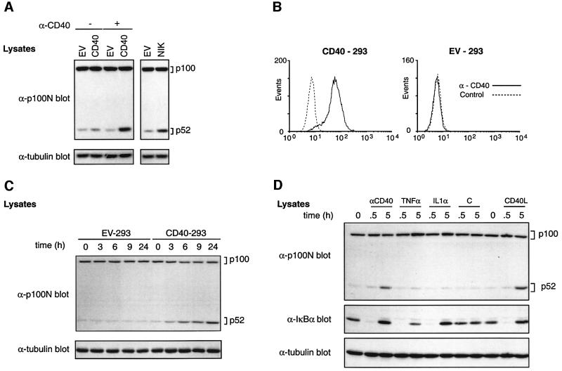
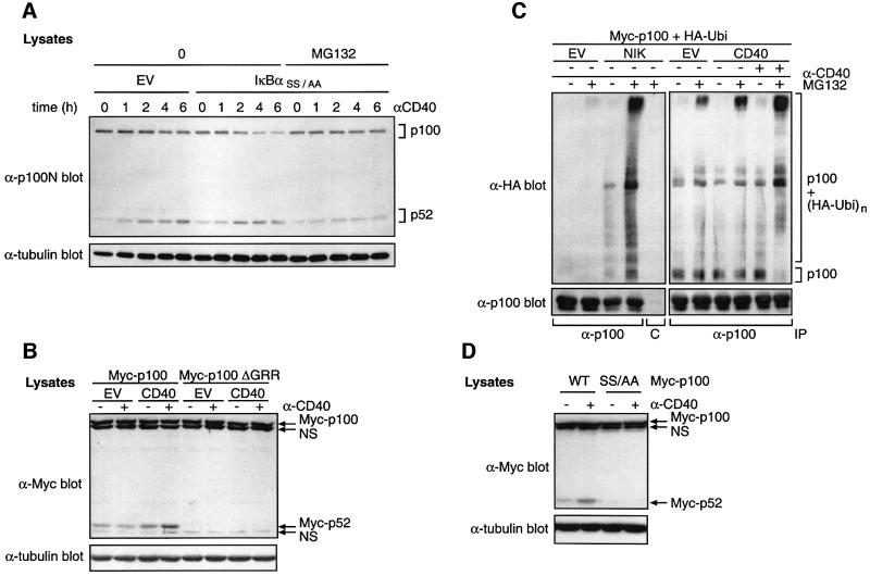
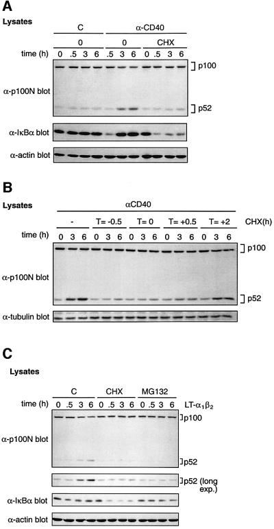
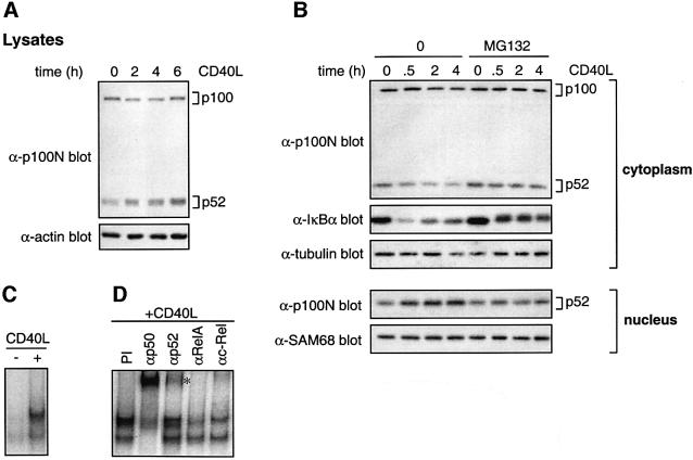
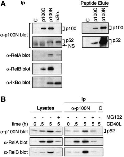
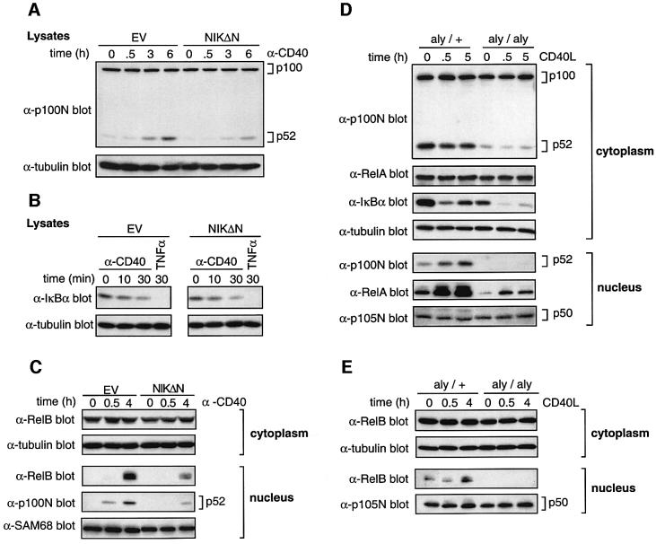
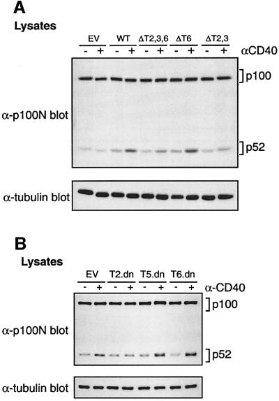
The nf-kb2 gene encodes the cytoplasmic NF-kappaB inhibitory protein p100 from which the active p52 NF-kappaB subunit is derived by proteasome-mediated proteolysis. Ligands which stimulate p100 processing to p52 have not been defined. Here, ligation of CD40 on transfected 293 cells is shown to trigger p52 production by stimulating p100 ubiquitylation and subsequent proteasome-mediated proteolysis. CD40-mediated p52 accumulation is dependent on de novo protein synthesis and triggers p52 translocation into the nucleus to generate active NF-kappaB dimers. Endogenous CD40 ligation on primary murine splenic B cells also stimulates p100 processing, which results in the delayed nuclear translocation of p52-RelB dimers. In both 293 cells and primary splenic B cells, the ability of CD40 to trigger p100 processing requires functional NF-kappaB-inducing kinase (NIK). In contrast, NIK activity is not required for CD40 to stimulate the degradation of IkappaBalpha in either cell type. The regulation of p100 processing by CD40 is likely to be important for the transcriptional regulation of CD40 target genes in adaptive immune responses.


Figures

None
Fig. 1. CD40 induces p52 production in transfected 293 cells. (A) 293 cells were transfected with the indicated vectors. After 24 h, anti-CD40 (+) or control (–) mAbs were added and cells incubated for a further 14 h. Cell lysates were western blotted. (B) 293 cells were transfected with a plasmid encoding CD40 or with empty vector (EV), and clones were isolated after G418 selection. CD40 expression of a representative CD40-transfected clone (CD40-293, clone B4) or EV-transfected 293 cells (EV-293) was determined by flow cytometry. (C) CD40-293 or EV-293 cells were stimulated for the indicated times with anti-CD40 mAb (10 µg/ml) and lysates western blotted. Similar kinetics of p52 induction were detected over a range of anti-CD40 mAb concentrations (see Supplementary figure 1 available at The EMBO Journal Online). (D) CD40-293 cells were stimulated with anti-CD40 mAb, TNF-α, IL-1α or CD40L for the indicated times and lysates western blotted.
None
Fig. 2. CD40 induces ubiquitylation and proteasome-mediated processing of p100 to generate p52. (A) CD40-293 cells were transfected with a plasmid encoding IκBαSS/AA or with no insert as a control. Cells were cultured for 24 h, pre-cultured for 30 min with MG132 or control vehicle and then stimulated for the indicated times with anti-CD40 or left unstimulated. Western blots of cell lysates were probed as shown. IκBαSS/AA was expressed at similar levels in all transfections as determined by western blot analysis (data not shown). (B) 293 cells were co-transfected with plasmids encoding CD40 or EV and Myc-p100 or Myc-p100ΔGRR. Cells were stimulated with anti-CD40 mAb for the last 15 h of a 48 h culture and lysates western blotted. The positions of non-specific bands (NS) are indicated. (C) 293 cells were co-transfected with plasmids encoding Myc-p100 and HA-ubiquitin (HA-Ubi) together with plasmids encoding CD40, NIK or with no insert (EV). After 48 h culture, cells were incubated for 15 min with 50 µM MG132 and then stimulated for 1 h with anti-CD40 or control mAb. p100 was immunoprecipitated from cell lysates with anti-hp100 antibody and then resolved on a 6% SDS–polyacrylamide gel and western blotted. Pre-immune serum (C) was used a as a control for immunoprecipitations. (D) 293 cells were co-transfected with vectors encoding CD40 and Myc-p100 or Myc-p100(S866A,S870A) (SS/AA). Cells were incubated with anti-CD40 or control mAb for the last 15 h of a 48 h culture period and lysates western blotted. Similar levels of CD40 expression in each transfection were confirmed by immunoblotting with anti-CD40 antibody (data not shown).
None
Fig. 3. CD40 induction of p52 is dependent on de novo protein synthesis. (A) CD40-293 cells were pre-treated with cycloheximide (CHX) or vehicle control (0) for 30 min. Cells were then stimulated with anti-CD40 or control (C) mAb for the indicated times and lysates western blotted. (B) CHX was added to cultures of CD40-293 cells at the indicated times relative to anti-CD40 mAb. Cell lysates were western blotted. (C) 293 cells were pre-treated for 30 min with MG132, CHX or control medium (C). Cells were then stimulated for the times shown with LTα1β2, and lysates western blotted.
None
Fig. 4. CD40 ligation induces nuclear translocation of p52 dimers. (A) Nuclear and cytoplasmic fractions were prepared from CD40-293 cells stimulated as indicated and western blotted. (B) Nuclear fractions were prepared from CD40-293 cells stimulated as shown. NF-κB DNA-binding activity was analysed by EMSA. (C) EMSAs were carried out on nuclear extracts from CD40-293 cells stimulated for 0.5 or 6 h with anti-CD40 mAb. Extracts were supershifted with the indicated antibodies to different Rel proteins or pre-immune serum (PI). The position of a p52-containing supershifted NF-κB complex is shown (asterisk). Specificity of κB binding was determined by competition with 100-fold unlabelled κB oligonucleotide (Oligo, NF-κB) or control Oct-1 oligonucleotide (Oligo, Oct-1). (D) Nuclear extracts from CD40-293 cells were supershifted with anti-p100N antibody and analysed by EMSA. The position of supershifted complexes is shown (arrow).
None
Fig. 5. CD40 ligation induces p100 proteolysis and nuclear translocation of p52 in splenic B cells. (A and B) Splenic B cells were stimulated in vitro with CD40L for the times shown or left unstimulated. Cell lysates (A) or cytoplasmic and nuclear fractions (B) were western blotted. (C) NF-κB EMSAs were carried out on nuclear fractions from primary splenic B cells stimulated with CD40L for 5 h or left unstimulated. (D) Nuclear extracts prepared from splenic B cells stimulated for 5 h with CD40L were supershifted with the indicated anti-Rel antibodies or pre-immune serum (PI). The position of the p52-containing supershifted NF-κB complex is shown (asterisk).
None
Fig. 6. CD40 induces nuclear accumulation of p52–RelA and p52– RelB dimers in splenic B cells. (A) Lysates of unstimulated splenic B cells were immunoprecipitated with the indicated antibodies and western blotted. In the right hand panels, specific peptide was used to elute bound antigen to decrease background Ig. (B) Nuclear extracts were prepared from splenic B cells stimulated with CD40L for 0.5 or 5 h or left unstimulated. MG132 was added 15 min prior to stimulation. p52 was then immunoprecipitated and associated Rel subunits identified by western blotting. C indicates control IgG immunoprecipitations.
None
Fig. 7. CD40 stimulation of p100 processing requires NIK activity. (AC) CD40-293 cells were transfected with plasmids encoding Myc-NIKΔN (NIKΔN) or with no insert as a control (EV). Cells were stimulated as indicated. Lysates (A and B) or cytoplasmic and nuclear fractions (C) were western blotted. Equivalent expression of Myc-NIKΔN was confirmed by western blotting (data not shown). (D and E) Splenic B cells were isolated from aly/aly and aly/+ mice. Cells were stimulated with CD40L for 0.5 or 4 or 5 h, or left unstimulated (0). Cytoplasmic and nuclear fractions were western blotted.
None
Fig. 8. Efficient p52 induction by CD40 requires its TRAF2/3-binding site. (A) 293 cells were transiently transfected with vectors encoding the indicated CD40 chimeras. Cells were stimulated with anti-CD40 mAb for the last 16 h of a 48 h culture and lysates western blotted. Each of the CD40 chimeras was expressed at similar levels (data not shown). (B) 293 cells were co-transfected with vectors encoding CD40 and dominant-negative TRAFs or EV. Cells were stimulated for the last 16 h of a 48 h culture and lysates western blotted. Similar levels of CD40 were expressed in all transfections, and each of the TRAF dominant-negative proteins was expressed at comparable levels (data not shown).

References

    1. Ahonen C.L., Manning,E.M., Erickson,L.D., O’Connor,B.P., Lind,E.F., Pullen,S.S., Kehry,M.R. and Noelle,R.J. (2002) The CD40–TRAF6 axis controls affinity maturation and the generation of long-lived plasma cells. Nat. Immunol., 3, 451–456. - PMC - PubMed
    1. Aizawa S. et al. (1997) Tumor necrosis factor receptor-associated factor (TRAF) 5 and TRAF2 are involved in CD30-mediated NF-κB activation. J. Biol. Chem., 272, 2042–2045. - PubMed
    1. Alkalay I., Yaron,A., Hatzubai,A., Jung,S., Avraham,A., Gerlitz,O., Pashut-Lavon,I. and Ben-Neriah,Y. (1995) In vivo stimulation of IκB phosphorylation is not sufficient to activate NF-κB. Mol. Cell. Biol., 15, 1294–1301. - PMC - PubMed
    1. Baeuerle P.A. and Henkel,T. (1994) Function and activation of NF-κB in the immune system. Annu. Rev. Immunol., 12, 141–179. - PubMed
    1. Beinke S., Belich,M.P. and Ley,S.C. (2002) The death domain of NF-κB1 p105 is essential for signal-induced p105 proteolysis. J. Biol. Chem., 277, 24162–24168. - PubMed

Publication types

MeSH terms

Substances