. 2002 Oct 15;21(20):5396-407.
doi: 10.1093/emboj/cdf551.

A phosphoserine/threonine-binding pocket in AGC kinases and PDK1 mediates activation by hydrophobic motif phosphorylation

Affiliations

A phosphoserine/threonine-binding pocket in AGC kinases and PDK1 mediates activation by hydrophobic motif phosphorylation

Morten Frödin et al. EMBO J. .

Abstract

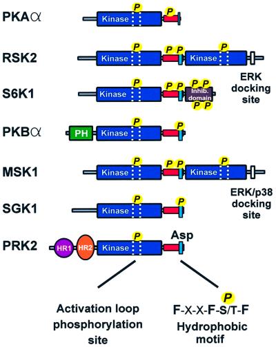
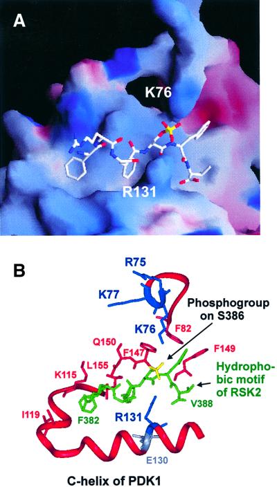
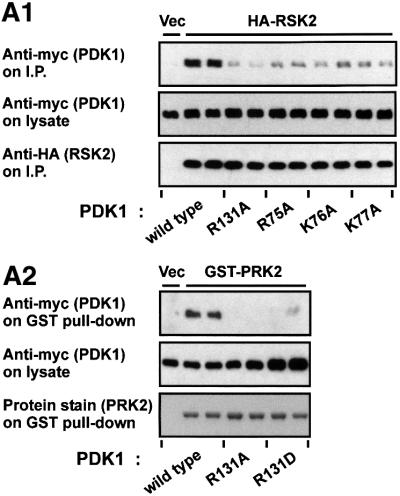
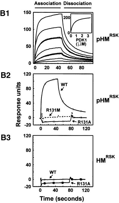
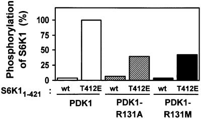
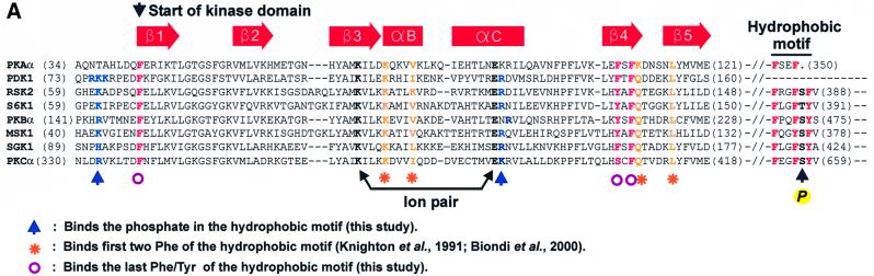
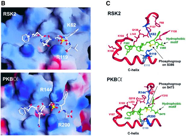
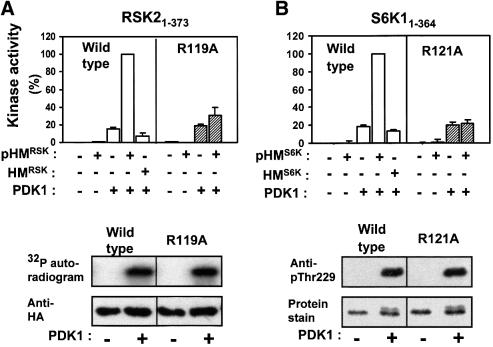
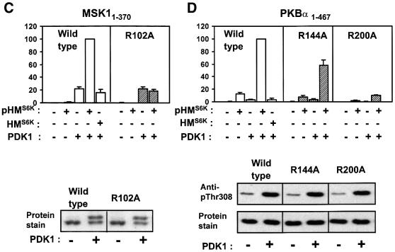
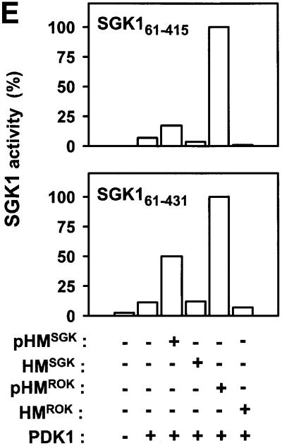
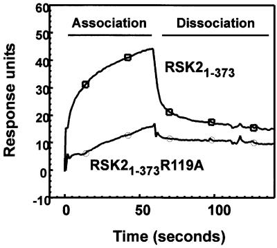
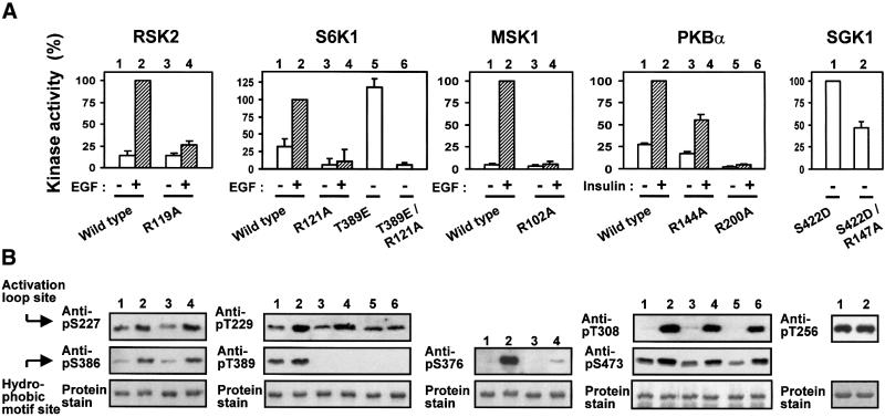
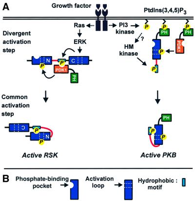
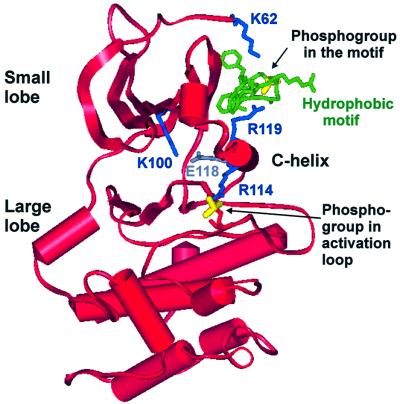
The growth factor-activated AGC protein kinases RSK, S6K, PKB, MSK and SGK are activated by serine/threonine phosphorylation in the activation loop and in the hydrophobic motif, C-terminal to the kinase domain. In some of these kinases, phosphorylation of the hydrophobic motif creates a specific docking site that recruits and activates PDK1, which then phosphorylates the activation loop. Here, we discover a pocket in the kinase domain of PDK1 that recognizes the phosphoserine/phosphothreonine in the hydrophobic motif by identifying two oppositely positioned arginine and lysine residues that bind the phosphate. Moreover, we demonstrate that RSK2, S6K1, PKBalpha, MSK1 and SGK1 contain a similar phosphate-binding pocket, which they use for intramolecular interaction with their own phosphorylated hydrophobic motif. Molecular modelling and experimental data provide evidence for a common activation mechanism in which the phosphorylated hydrophobic motif and activation loop act on the alphaC-helix of the kinase structure to induce synergistic stimulation of catalytic activity. Sequence conservation suggests that this mechanism is a key feature in activation of >40 human AGC kinases.


Figures

None
Fig. 1. Structural alignment of PKA and major growth factor-activated AGC kinases. The alignment illustrates that the growth factor-activated AGC kinases share two regulatory features: phosphorylation of the activation loop (stippled area) and phosphorylation of a hydrophobic motif (blue box), located in a tail region (red box) C-terminal to the kinase domain. Note that PRK2 contains a phosphate-mimicking aspartic acid residue and that PKA lacks a phosphorylation site in the hydrophobic motif.
None
Fig. 2. Model of the docking interaction between PDK1 and the phosphorylated hydrophobic motif of RSK2. (A) Electrostatic surface potential model of the hydrophobic pocket of PDK1 with positive potential in blue and negative potential in red. The RSK2 hydrophobic motif peptide (FRGFpSFV) is shown in white with the phosphogroup on Ser386 in yellow. (B) Ribbon representation of the pocket with side chains of the residues discussed in the text. PDK1 residues are red, blue or grey, whereas the RSK2 hydrophobic motif peptide is shown in green with the phosphogroup in yellow.
None
Fig. 3. Identification of arginine/lysine residues in PDK1 required for interaction with the phosphorylated hydrophobic motif. (A) COS7 cells were co-transfected with plasmids expressing wild-type or mutant myc-PDK1 together with HA-RSK2, GST–PRK2 or empty vector (Vec). After 48 h and a final 3 h serum starvation period, the cells were lysed subsequent to 35 min EGF treatment of RSK-expressing cells. HA-RSK2 and GST–PRK2 were precipitated from the cell lysates using anti-HA antibody or glutathione beads, respectively. The precipitates were subjected to SDS–PAGE and immunoblotting with anti-myc antibody to detect co-precipitated myc-PDK1 (upper panels) or to anti-HA immunostaining or protein staining to assess the amounts of HA-RSK2 and GST–PRK2 (lower panels). Pre-precipitation lysates were subjected to immunoblotting for the myc tag (middle panels). (B) Interaction of PDK1 with the hydrophobic motif of RSK2 was analysed using surface plasmon resonance measurements in a BiaCore3000 system. Biotinylated peptides of the motif phosphorylated (pHMRSK) or non-phosphorylated (HMRSK) at Ser386 were used to coat Sensor Chips SA (10 response units). (1) GST–PDK1 was injected at different concentrations (0.013–3.33 µM) onto pHM-coated chips. In the inset, the steady-state binding is plotted against the various concentrations of PDK1. Kinetic constants were obtained by fitting the data to a hyperbola using Kaleidagraph software. (2 and 3) GST–PDK1 wild-type, –R131A or –R131M were injected at 400 nM onto chips coated with pHMRSK and HMRSK, respectively. All experiments in (A) and (B) were performed three times with similar results.
None
Fig. 4. R131 is important for the ability of PDK1 to phosphorylate S6K1. Wild-type or T412E His-S6K11–421 were incubated for 10 min with wild-type or mutant GST–PDK1 and Mg[γ-32P]ATP. Thereafter, the kinase reactions were subjected to SDS–PAGE. Radioactivity incorporated into the S6K1 protein band was quantitated and expressed as a percentage of the maximal value obtained. The results are representative of two independent experiments.
None
Fig. 5. A phosphate-binding pocket is conserved in major growth factor-activated AGC kinases. (A) Amino acid sequence alignment of selected AGC kinases in the region of the kinase domain that contains the hydrophobic pocket as well as the region forming the hydrophobic motif. Conserved residues predicted to interact with the phosphogroup, the first two aromatic residues and the last aromatic residue of the hydrophobic motif are indicated by an arrow, asterisk and circle, respectively. The ion pair formation between the lysine and glutamic acid residues conserved in all kinases is indicated. (B and C) Model of the intramolecular interaction of the hydrophobic pocket of RSK2 and PKBα with their respective phosphorylated hydrophobic motifs, FRGFpSFV (RSK2) and FPQFpSYS (PKBα). (B) Electrostatic surface potential models of the pockets, with positive potential in blue and negative potential in red. The hydrophobic motif peptides are shown in white with the phosphogroup in yellow. (C) Ribbon representation of the pockets with side chains of the residues discussed in the text. The hydrophobic motif peptides are shown in green with the phosphogroup in yellow.
None
Fig. 6. Evidence for intramolecular activation of AGC kinases by the phosphorylated hydrophobic motif. (AE) Upper panels: wild-type or point-mutated kinase domains of RSK2, S6K1, MSK1, PKBα and SGK1, all lacking their hydrophobic motif (except SGK161–431), were pre-phosphorylated or not by PDK1. Thereafter, PDK1 was removed (except in the RSK2 assay). The activity of each kinase was then determined in the absence or presence of phosphorylated (pHM) or non-phosphorylated (HM) hydrophobic motif peptide at 170 µM (A, B and E) or 360 µM (C and D) and expressed as a percentage of maximal activity. Data are means ± SD of (A) 4–10 observations from 2–4 independent experiments, (B–D) three independent experiments or (E) means of duplicates with <5% difference between duplicate samples from one experiment performed twice with similar results (A–D, lower panels). To control for an equal amount of protein and PDK1-induced phosphorylation of wild-type and mutant kinase, aliquots of the reactions were subjected to SDS–PAGE and protein staining/anti-HA blotting or to blotting with phospho-specific antibody to the PDK1 site. However, in (A), phosphorylation of HA-RSK21–373 was assessed by including [γ-32P]ATP during pre-incubation with PDK1, followed by precipitation with anti-HA antibody, SDS–PAGE and autoradiography.
None
Fig. 7. Interaction of RSK2 with its phosphorylated hydrophobic motif. The interaction of RSK2 with its own hydrophobic motif was analysed by surface plasmon resonance measurements. Biotinylated pHMRSK peptide was used to coat Sensor Chips SA (500 response units) and tested for binding to GST–HA-RSK21–373 or GST–HA-RSK21–373 R119A. The experiments were performed several times with similar results.
None
Fig. 8. Role of the phosphate-binding arginines in activation and phosphorylation of AGC kinases in vivo. COS7 cells were transfected with plasmids expressing HA- or GST-tagged wild-type or mutant kinase. After 48 h and a final 4 h serum starvation period, cells were exposed to 20 nM EGF for 20 min or 1 µM insulin for 8 min as indicated, and then lysed. Thereafter, the kinases were precipitated from the cell lysates with antibody to the HA tag or with glutathione beads. (A) Kinase activity was determined and expressed as a percentage of the wild-type stimulated value. Data are means ± SD of three (RSK2, S6K1, MSK1 and SGK1) or four (PKBα) independent experiments performed in duplicate. (B) Precipitated kinases from (A) were subjected to SDS–PAGE. The gel was subjected to immunoblotting with the indicated anti-phosphopeptide antibody or stained for protein.
None
Fig. 9. Roles of the phosphate-binding pocket in regulation of the growth factor-activated AGC kinases. (A) The activation mechanism of AGC kinases may be divided into a divergent and a common activation step. In both steps, phosphoserine/phosphothreonine recognition by the phosphate-binding pocket may play a key role, as exemplified here with RSK and PKB. (1) In the divergent step, the kinases are subject to pathway-specific regulation via signalling modules flanking the kinase domain and which leads to phosphorylation of the hydrophobic motif (in RSK by the C-terminal kinase domain and in PKB by an as yet unknown kinase). In RSK (and S6K or SGK), the phosphate of the hydrophobic motif is then recognized by the phosphate-binding pocket of PDK1, resulting in recruitment of PDK1 and subsequent phosphorylation of the activation loop. Thus, the regulatory phosphorylations occur in a mandatory order. In PKB, PDK1 is recruited via PH domain-mediated co-localization at the cell membrane, and thus the order of the regulatory phosphorylations does not appear mandatory. (2) The second activation step may be common to all AGC kinases that contain a phosphorylatable hydrophobic motif, thus also PKC, Rho kinase and Ndr not studied here. In this step, the phosphate-binding site promotes intramolecular binding of the hydrophobic motif to the hydrophobic pocket within the AGC kinase domain. In concert with activation loop phosphorylation, this brings about a conformational change in the kinase domain, leading to catalytic activation. (B) Key to major symbols used in (A).
None
Fig. 10. Model of how the phosphates of the hydrophobic motif and the activation loop cooperate to activate AGC kinases. Ribbon representation of the model in Figure 5B of RSK2 interacting with its hydrophobic motif, but viewed from another angle and showing the entire RSK2 kinase domain. The figure illustrates how the phosphates of the hydrophobic motif and the activation loop can cooperate to position the C-helix for optimal ion pairing of Glu118 and the ATP-binding Lys100. Moreover, the C-helix, fixed by the hydrophobic motif, can function as a bridge between the small and large lobes to stabilize the kinase in the closed (active) conformation, which is shown here.

References

    1. Alessi D.R., Andjelkovic,M., Caudwell,B., Cron,P., Morrice,N., Cohen,P. and Hemmings,B.A. (1996) Mechanism of activation of protein kinase B by insulin and IGF-1. EMBO J., 15, 6541–6551. - PMC - PubMed
    1. Alessi D.R., James,S.R., Downes,C.P., Holmes,A.B., Gaffney,P.R., Reese,C.B. and Cohen,P. (1997) Characterization of a 3-phospho inositide-dependent protein kinase which phosphorylates and activates protein kinase Bα. Curr. Biol., 7, 261–269. - PubMed
    1. Alessi D.R., Kozlowski,M.T., Weng,Q.P., Morrice,N. and Avruch,J. (1998) 3-phosphoinositide-dependent protein kinase 1 (PDK1) phosphorylates and activates the p70 S6 kinase in vivo and in vitro. Curr. Biol., 8, 69–81. - PubMed
    1. Balendran A., Biondi,R.M., Cheung,P.C., Casamayor,A., Deak,M. and Alessi,D.R. (2000) A 3-phosphoinositide-dependent protein kinase-1 (PDK1) docking site is required for the phosphorylation of protein kinase Cζ (PKCζ) and PKC-related kinase 2 by PDK1. J. Biol. Chem., 275, 20806–20813. - PubMed
    1. Biondi R.M., Cheung,P.C., Casamayor,A., Deak,M., Currie,R.A. and Alessi,D.R. (2000) Identification of a pocket in the PDK1 kinase domain that interacts with PIF and the C-terminal residues of PKA. EMBO J., 19, 979–988. - PMC - PubMed

Publication types

MeSH terms

Substances