. 2002 Oct 15;21(20):5516-26.
doi: 10.1093/emboj/cdf555.

Pre-spliceosome formation in S.pombe requires a stable complex of SF1-U2AF(59)-U2AF(23)

Affiliations

Pre-spliceosome formation in S.pombe requires a stable complex of SF1-U2AF(59)-U2AF(23)

Tao Huang et al. EMBO J. .

Abstract

We have initiated a biochemical analysis of splicing complexes in extracts from the fission yeast Schizosaccharomyces pombe. Extracts of S.pombe contain high levels of the spliceosome-like U2/5/6 tri-snRNP, which dissociates into mono-snRNPs in the presence of ATP, and supports binding of U2 snRNP to the 3' end of introns, yielding a weak ATP-independent E complex and the stable ATP-dependent complex A. The requirements for S.pombe complex A formation (pre-mRNA sequence elements, protein splicing factors, SF1/BBP and both subunits of U2AF) are analogous to those of mammalian complex A. The S.pombe SF1/BBP, U2AF(59) and U2AF(23) are tightly associated in a novel complex that is required for complex A formation. This pre-formed SF1- U2AF(59)-U2AF(23) complex may represent a streamlined mechanism for recognition of the branch site, pyrimidine tract and 3' splice site at the 3' end of introns.


Figures
None
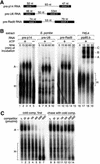
Fig. 1. Extracts of S.pombe efficiently form a complex on S.pombe pre-mRNAs. (A) Schematic of S.pombe pre-mRNA constructs used in this study. Pre-p14 is derived from the gene for the U2 snRNP p14 component (Will et al., 2001; SPBC29A3.07c), pre-U6 from the gene for U6 snRNA (X14196, M55650) and pre-Rad9 from the gene for DNA repair protein Rad9 (SPAC664.07c). Solid boxes represent exons, and lines represent introns. (B) Formation of complexes on pre-p14 RNA (lanes 1–5), pre-U6 RNA (lanes 6–10) and pre-Rad9 RNA (lanes 11–15) in S.pombe extract, or for comparison on adenovirus-derived pre-mRNA in a HeLa cell nuclear extract (lanes 16–19). RNAs were incubated in extract at 30°C for the times indicated, adjusted to 0.5 mg of heparin/ml, separated on a native 4% polyacrylamide gel and visualized by phosphoimaging. A, U2 snRNP complexes containing pre-mRNA; B, spliceosomal complex B containing U2/4/5/6 snRNPs and pre-mRNA; C, spliceosomal complex C containing U2/5/6 snRNPs and splicing intermediates; H, non-specific complexes. (C) Stability of S.pombe complex A. Unlabeled competitor pre-p14 RNA was added either before (lanes 1–7) or after (lanes 8–14) formation of complex A on labeled pre-p14 RNA and reincubated at 30°C for 30 min. Complexes were analyzed as in (B).
None
Fig. 2. The complex formed in extracts contains U2 snRNP, analogously to complex A. (A) snRNA requirement. Formation of complexes on pre-p14 RNA using extracts in which individual snRNAs were targeted for degradation by incubation with an antisense DNA oligonucleotide and RNase H. Untreated extract (lanes 1–3); mock-treated extract (lanes 4–6 and 13–15); extracts treated with oligonucleotides complementary to the branch site pairing region of U2 snRNA (nucleotides 28–42; lanes 7–9), U6 snRNA (nucleotides 21–69; lanes 10–12) or the 5′ end of U1 snRNA (nucleotides 1–14; lanes 16–18). Complexes were analyzed as in Figure 1B. (B) Native northern blot analysis of the complex. Extract was incubated with or without 400 pmol/ml unlabeled pre-p14 RNA at 30°C for 30 min (with ATP), separated on a native gel and transferred to Hybond N membrane, which was probed sequentially using riboprobes complementary to U2, U1, U4, U5 and U6 snRNAs. (C) Northern blot analysis of targeted snRNAs. Aliquots of extracts used in (A) were deproteinized, separated by 10% PAGE, blotted to Nytran and hybridized with antisense oligonucleotides to assess the extent of targeted degradation. (D) snRNA composition. Biotinylated pre-p14 RNA was incubated in extracts as indicated above each lane, bound to streptavidin–agarose beads and washed. Bound complexes were eluted and the RNAs separated and probed as in (C).
None
Fig. 3. Analysis of S.pombe snRNP distribution: S.pombe extracts contain U2/5/6 tri-snRNPs and few U4/5/6 tri-snRNPs. (A) Dissociation of U2/5/6 upon incubation with ATP, and accumulation of U2 in complex A. Native northern blot analysis of the complex. Extract was incubated with or without ATP and with or without 400 pmol/ml unlabeled pre-U6 RNA at 30°C for 30 min as indicated, separated on a native gel and transferred to Hybond N membrane. The membrane was probed using a riboprobe complementary to U2 snRNA. (B) Glycerol gradient fractionation and subsequent native gel separation of extracts from wild-type cells. Extract was fractionated through a 10–30% glycerol gradient; fractions were loaded directly onto a native gel, which was blotted and probed consecutively for each snRNA as indicated. The positions of 40S and 60S ribosomal subunits from parallel gradients are indicated. (C) Analysis as in (B) of U2AF59ts mutant extract, after growth at the non-permissive temperature for 2 h.
None
Fig. 4. Sequence elements required for S.pombe complex A formation. (A) Schematic of pre-p14 constructs. SS, splice site; BS, branch site; Py tract, pyrimidine tract. (B) Formation of complexes on mutant pre-p14 RNAs. RNAs represented in (A) were incubated in S.pombe extract at 30°C for the times indicated, and separated on a native gel, as in Figure 1B. (C) Formation of complexes on the 3′ half pre-p14 RNA using extracts in which individual snRNAs were targeted for degradation by RNase H as in Figure 2A. (D) Formation of complexes on 3′ half pre-p14 RNAs containing 3′SS or Py tract mutations.
None
Fig. 5. The S.pombe complex A requires U2AF. (A) Formation of complex A on pre-p14 RNA in extracts from a U2AF ts mutant (prp2.2; Beales et al., 2000), grown at permissive temperature (lanes 1–7) or shifted for 2 h to the non-permissive temperature (lanes 8–14). Extracts were pre-incubated for 30 min either on ice (lanes 1, 2, 8 and 9) or at 37°C to heat inactivate (lanes 3–7 and 10–14). Lanes 5 and 12, addition of U2AF-TAP purified complexes; lanes 6 and 13, addition of mock-purified material; lanes 7 and 14, addition of Prp43-TAP purified material. Complexes were analyzed as in Figure 1B. (B) Schematic of TAP fusion proteins. CBP, calmodulin-binding peptide; TEV, TEV protease cleavage site; protein A, two copies of the IgG-binding domain derived from protein A (Puig et al., 2001). (C) Extent of tagged protein depletion. Extracts containing TAP-tagged SF1 (SPCC962.06c), U2AF59 (SPBC146.07), Prp43p (SPBC16H5.10c) or Prp5p (SPCC10H11.01) (lanes 2, 4, 6 and 8) were depleted of tagged proteins using IgG–Sepharose (lanes 3, 5, 7 and 9), separated on a 10% polyacrylamide gel, western blotted and probed for the TAP tag. WT, extract from untagged wild-type cells. Control, probed with antiserum to spindle checkpoint protein Slp1 as a loading control. (D) Depletion of U2AF and SF1/BBP and reconstitution of complex A. Formation of complexes on pre-p14 RNA using extracts in which various proteins had been TAP tagged, depleted and supplemented with partially purified protein complexes. Wild-type untagged extract (lanes 1 and 2), extracts in which Prp43p, SF1 or U2AF59 were TAP tagged (lanes 3 and 4, 5 and 6, and 7 and 8, respectively); extract that was mock-treated under depletion conditions (lanes 9 and 10), extracts depleted of Prp43p-TAP, SF1-TAP, U2AF59-TAP and Prp5p-TAP (lanes 11 and 12, 13 and 14, 15 and 16, and 17 and 18, respectively); mixture of SF1-depleted and U2AF59-depleted extracts (lanes 19 and 20); mixture of SF1-depleted and Prp5p-depleted extracts (lanes 21 and 22); mixture of U2AF59-depleted and Prp5p-depleted extracts (lanes 23 and 24). Lanes 25–32: SF1-depleted extract or U2AF59-depleted extract was supplemented with mock-purified, SF1-purified, U2AF59-purified or Prp43p-purified fractions.
None
Fig. 6. U2AF and SF1/BBP are tightly complexed. (A) Affinity selection of the U2AF–SF1 complex. Lysates from double-tagged cells, containing TAP- and HA-tagged proteins as indicated, were incubated with IgG–Sepharose beads to bind the TAP moiety. Co-purifying proteins were analyzed by western blot using anti-HA antibodies. Lanes 1–6, half of the input lysate; lanes 7–12, HA-tagged proteins present in the purified material; lanes 13–18, half of the input and IgG-selected material from cells lacking any TAP-tagged protein as a control; 59, U2AF59; 23, U2AF23. (B) The U2AF–SF1 complex is stable to high salt. Lysate from triple-tagged cells (SF1-TAP, U2AF59-HA, U2AF23-HA) was incubated with IgG beads and washed at salt concentrations as indicated (mM). Affinity-selected proteins were analyzed as in (A). (C) The U2AF–SF1 complex is resistant to RNase. Lysate from triple-tagged cells as in (B) was incubated with 0.5 mg/ml RNase A for 30 min at 30°C prior to incubation with IgG beads. Affinity-selected proteins were analyzed as in (A). Lane 1, 1/15th of input lysate; lane 2, untreated lysate; lane 3, mock treatment; lane 4, lysates incubated with RNase.
None
Fig. 7. Schematic model of snRNP dynamics observed in S.pombe extract. U2/5/6 tri-snRNP undergoes an ATP-dependent dissociation, resulting in an increase in the apparent free pool of U2, U5 and U6. This free pool of U2 participates in weak binding to pre-mRNA in an ATP-independent E complex, which is dependent on SF1–U2AF59– U2AF23 complex. A second ATP-dependent step results in stable engagement of U2 on the pre-mRNA; the extent of concomitant displacement of SF1–U2AF59–U2AF23 is uncertain.

Similar articles

Cited by

References

    1. Abovich N. and Rosbash,M. (1997) Cross-intron bridging interactions in the yeast commitment complex are conserved in mammals. Cell, 89, 403–412. - PubMed
    1. Ansari A. and Schwer,B. (1995) SLU7 and a novel activity, SSF1, act during the PRP16-dependent step of yeast pre-mRNA splicing. EMBO J., 14, 4001–4009. - PMC - PubMed
    1. Arenas J.E. and Abelson,J.N. (1997) Prp43: an RNA helicase-like factor involved in spliceosome disassembly. Proc. Natl Acad. Sci. USA, 94, 11798–11802. - PMC - PubMed
    1. Bahler J., Wu,J.Q., Longtine,M.S., Shah,N.G., McKenzie,A.,III, Steever,A.B., Wach,A., Philippsen,P. and Pringle,J.R. (1998) Heterologous modules for efficient and versatile PCR-based gene targeting in Schizosaccharomyces pombe. Yeast, 14, 943–951. - PubMed
    1. Beales M., Flay,N., McKinney,R., Habara,Y., Ohshima,Y., Tani,T. and Potashkin,J. (2000) Mutations in the large subunit of U2AF disrupt pre-mRNA splicing, cell cycle progression and nuclear structure. Yeast, 16, 1001–1013. - PubMed

Publication types

MeSH terms