. 2002 Oct 15;21(20):5539-47.
doi: 10.1093/emboj/cdf547.

60S pre-ribosome formation viewed from assembly in the nucleolus until export to the cytoplasm

Affiliations

60S pre-ribosome formation viewed from assembly in the nucleolus until export to the cytoplasm

Tracy A Nissan et al. EMBO J. .

Abstract

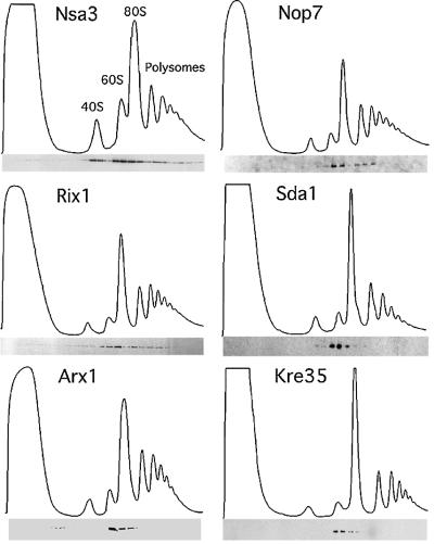
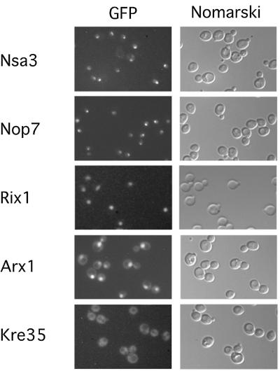
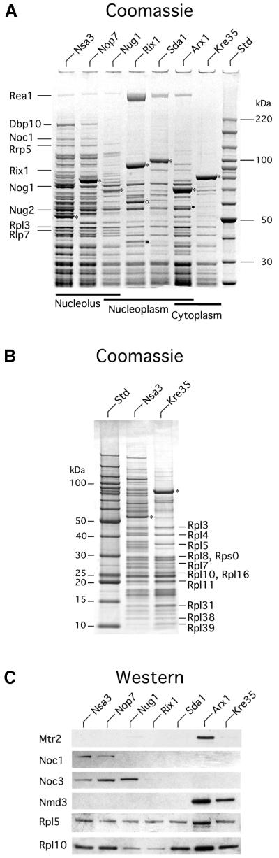
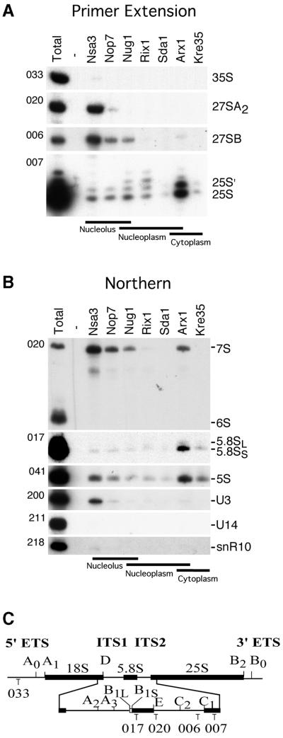
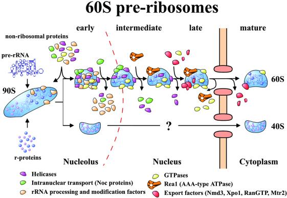
60S ribosomes undergo initial assembly in the nucleolus before export to the cytoplasm and recent analyses have identified several nucleolar pre-60S particles. To unravel the steps in the pathway of ribosome formation, we have purified the pre-60S ribosomes associated with proteins predicted to act at different stages as the pre-ribosomes transit from the nucleolus through the nucleoplasm and are then exported to the cytoplasm for final maturation. About 50 non-ribosomal proteins are associated with the early nucleolar pre-60S ribosomes. During subsequent maturation and transport to the nucleoplasm, many of these factors are removed, while others remain attached and additional factors transiently associate. When the 60S precursor particles are close to exit from the nucleus they associate with at least two export factors, Nmd3 and Mtr2. As the 60S pre-ribosome reaches the cytoplasm, almost all of the factors are dissociated. These data provide an initial biochemical map of 60S ribosomal subunit formation on its path from the nucleolus to the cytoplasm.


Figures

None
Fig. 1. Reverse-tagged protein baits of the Nug1-containing 60S pre- ribosome are associated with pre-ribosomal particles of different sizes. The sedimentation behavior of the indicated TAP-tagged proteins was analyzed on sucrose density gradients, and ribosomal fractions (40S, 60S, 80S and polysomes) were determined by OD254 measurement of the gradient fractions (upper graph). Western blot analysis of these gradient fractions using anti-ProtA antibodies reveals the position of the indicated TAP-tagged baits (lower panel). Note that some baits (e.g. Nsa3) exhibit a broad distribution on the sucrose gradient, whereas other baits (e.g. Kre35) exhibit a distinct peak at ∼60S (see text).
None
Fig. 2. Subcellular location of GFP-tagged protein baits in yeast cells. The in vivo location of the indicated tagged proteins, associated with different 60S pre-ribosomes, was analyzed by fluorescence microscopy. For microscopic inspection, cells were grown to mid-log phase, mounted on a microscope slide and photographed with identical exposure times.
None
Fig. 3. SDS–PAGE analysis of the protein composition of the different pre-60S ribosomes. (A) The indicated TAP-tagged protein baits were isolated from yeast lysates by a two-step affinity purification (TAP method), and TAP-purified bait proteins were separated on an 4–12% SDS–polyacrylamide gradient gel and stained with colloidal Coomassie Blue. Co-purifiying proteins were identified by mass spectrometry (MALDI-TOF) and prominent co-migrating bands are indicated. The positions of tagged proteins are indicated by asterisks, Nmd3 in the Arx1 preparation by a filled circle, and Ynl182p and Yhr085p in the Rix1 preparation by an open circle and closed square, respectively. The molecular weight marker is indicated on the right. Note the relative higher loading of the Arx1 particle in comparison with the Rix1, Sda1 and Kre35 particle loading. (B) Lower molecular weight proteins in earliest (Nsa3) and latest (Kre35) particles as visualized with 10–20% SDS–PAGE. All bands, both in the high (Table I) and low molecular weight range (Table II), were identified by MS. Each bait protein is indicated with an asterisk. In addition, selected ribosomal proteins are indicated. (C) Western blot analysis of the purified bait proteins, analyzed by SDS–PAGE as shown in (A) and listed in Supplementary table SI. The western blot was incubated with the indicated antibodies to Mtr2, Noc1, Noc3, Nmd3, Rpl5 and Rpl10. Not shown are Sqt1, which was identified in all purifications, and Tif6, which was present in all except Kre35.
None
Fig. 4. RNA analysis of the different pre-60S particles. (A) Primer extension and (B) northern hybridization analyses for 35S, 27S and 25S rRNA were performed on RNA extracted from whole cells (Total) and affinity-purified tagged (Nsa3, Nop7, Nug1, Rix1, Sda1, Arx1 and Kre35) and non-tagged (Negative) wild-type strain. RNA was loaded proportionally to a Coomassie stained protein SDS–PAGE from the same purifications. The oligonucleotide used for each panel is indicated on the left side of each gel (see Materials and methods). The annealing location of oligonucleotides used for primer extension and northern hybridization analysis are indicated (C).
None
Fig. 5. Model of the pathway of 60S pre-ribosome maturation and export. The maturation of the pre-ribosome is depicted. The various classes of factors that are associated with the particle are identified by color. The factors coming on and off the particles represent actual identified proteins by mass spectrometry. For simplicity, the alterations in RNA composition are not depicted.

References

    1. Adams C.C., Jakovljevic,J., Roman,J., Harnpicharnchai,P. and Woolford,J.L.,Jr (2002) Saccharomyces cerevisiae nucleolar protein Nop7p is necessary for biogenesis of 60S ribosomal subunits. RNA, 8, 150–165. - PMC - PubMed
    1. Baßler J., Grandi,P., Gadal,O., Leßmann,T., Tollervey,D., Lechner,J. and Hurt,E.C. (2001) Identification of a 60S pre-ribosomal particle that is closely linked to nuclear export. Mol. Cell, 8, 517–529. - PubMed
    1. Beltrame M. and Tollervey,D. (1992) Identification and functional analysis of two U3 binding sites on yeast pre-ribosomal RNA. EMBO J., 11, 1531–1542. - PMC - PubMed
    1. Buscemi G., Saracino,F., Masnada,D. and Carbone,M.L. (2000) The Saccharomyces cerevisiae SDA1 gene is required for actin cytoskeleton organization and cell cycle progression. J. Cell Sci., 113, 1199–1211. - PubMed
    1. Dragon F. et al. (2002) A large nucleolar U3 ribonucleoprotein required for 18S ribosomal RNA biogenesis. Nature, 417, 967–970. - PMC - PubMed

Publication types

MeSH terms