. 2002 Oct 15;11(22):2723-33.
doi: 10.1093/hmg/11.22.2723.

The inherited blindness associated protein AIPL1 interacts with the cell cycle regulator protein NUB1

Affiliations

The inherited blindness associated protein AIPL1 interacts with the cell cycle regulator protein NUB1

Dayna T Akey et al. Hum Mol Genet. .

Erratum in

  • Hum Mol Genet. 2003 Feb 15;12(4):451

Abstract

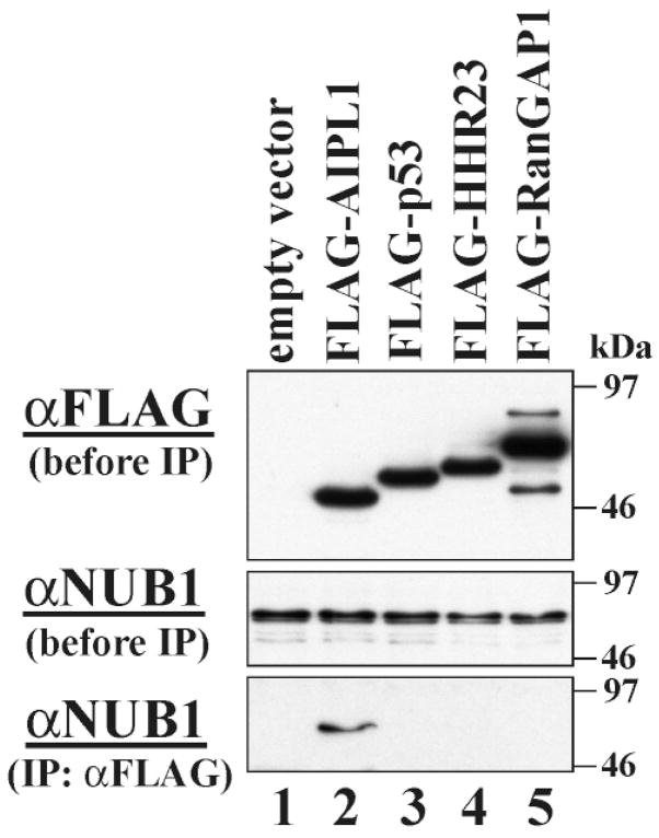
Mutations in the aryl hydrocarbon receptor-interacting protein-like 1 (AIPL1) gene have been found in patients with Leber congenital amaurosis (LCA), a severe, early-onset form of retinal degeneration. To determine the normal function of AIPL1 and to better understand how mutations in this gene cause disease, we performed a yeast two-hybrid screen to identify AIPL1-interacting proteins in the retina. One of the identified interacting proteins corresponds to NUB1 (NEDD8 Ultimate Buster 1), which is thought to control many biological events, especially cell cycle progression, by downregulating NEDD8 expression. The AIPL1-NUB1 interaction was verified by co-immunoprecipitation studies in Y79 retinoblastoma cells, demonstrating that this interaction occurs within cells that share a number of features with retinal progenitor cells. Furthermore, we examined the localization of the AIPL1 protein within developing and adult retinas, and found that AIPL1 is present in the developing photoreceptor layer of the human retina and within the photoreceptors of the adult retina. Similar to AIPL1, NUB1 is also expressed in the developing and adult retina. Therefore, it is possible that the early-onset form of retinal degeneration seen in LCA patients with AIPL1 mutations may be due to a defect in the regulation of cell cycle progression during photoreceptor maturation. These data raise the possibility that AIPL1 is important for appropriate photoreceptor formation during development and/or survival following differentiation.


Figures

Figure 1
Figure 1
Results of yeast two-hybrid retest with bovine Nub1. The pGAD10 plasmid containing bovine Nub1 was transformed into yeast strain AH109 with either the pGBKT7–bovine Aipl1 plasmid or the pGBKT7–empty plasmid. Transformants were grown on two different types of plates: (i) plates containing synthetic dropout media lacking Leu and Trp (−L/−W) to confirm transformation and (ii) plates containing dropout media with X-α-galactosidase (X-α-gal) and 3-AT (5 mM) but lacking Leu, Trp, and His (−L/−W/−H/+X-α-gal/+3AT) to assay for activation of the HIS3 and MEL1 reporters. (A) Transformation with pGBKT7–Aipl1 and pGAD10–Nub1 plasmids grown on −L/−W plates demonstrate that the transformation worked. (B) Transformation with pGBKT7–empty and pGAD10–Nub1 plasmids grown on −L/−W plates demonstrate that the transformation was successful. (C) Transformation with pGBKT7–Aipl1 and PGAD10–Nub1 plasmids. Formation of blue colonies on −L/−W/−H/+X-α-gal/+3AT plates indicates activation of MEL1 and HIS3 reporters and therefore the presence of a protein–protein interaction between AIPL1 and NUB1. (D) Transformation with pGBKT7–empty and pGAD10–bovine Nub1 plasmids. Lack of colony formation on −L/−W/−H/+X-α-gal/+3AT plates confirms that NUB1 requires the presence of AIPL1 to activate transcription of the HIS and MEL1 reporters.
Figure 2
Figure 2
Analysis of interaction in yeast. The pGADT7–AIPL1 deletion or mutant constructs shown on the left were co-transformed with the construct pGADT7–NUB1, and the activation of the HIS3 and LacZ reporter genes was determined (right) by a liquid growth assay (white bars) and a β-galactosidase assay (black bars), respectively.
Figure 3
Figure 3
Co-immunoprecipitation of endogenous NUB1 with FLAG–AIPL1. Empty vector (lane 1) or plasmid DNA encoding FLAG-tagged AIPL1 (lane 2), p53 (lane 3), HHR23 (lane 4) or RanGAP1 (lane 5) was overexpressed in COS cells. The total cell lysate was analyzed by antibody against FLAG (top panel) or NUB1 (middle panel). The total cell lysate was also incubated with anti-FLAG antibody for immunoprecipitation. Co-precipitated proteins were analyzed by immunoblotting using anti-NUB1 antibody (bottom panel). Only FLAG-tagged AIPL1 is capable of immunoprecipitating NUB1 (bottom panel, lane2), thereby demonstrating that these two proteins interact in vitro and in vivo. Molecular mass standards are expressed in kilodaltons.
Figure 4
Figure 4
Co-immunoprecipitation of NUB1 with AIPL1 in human Y79 retinoblastoma cells. Immunoprecipitates with preimmune serum (lane 1), anti-AIPL1 serum (lane 2) and affinity-purified anti-AIPL1 antibody (lane 3) were separated by SDS–PAGE and transferred for immunoblot analysis (IB) with either anti-NUB1 (A) or unpurified anti-AIPLl (B). Total Y79 cell lysate (lane 4) was also loaded on the gel.
Figure 5
Figure 5
Expression of Nub1 in the embryonic and postnatal mouse retina. Mouse Nub1 development in situ. Digoxygenin in situ hybridization of Nub1 mRNA is shown from embryonic day12 (E12) through to postnatal day 16 (P16). pcl, progenitor cell layer; gcl, ganglion cell layer; inl, inner nuclear layer; onl, outer nuclear layer; is, photoreceptor inner segments; P16 sense, sense control for background. Photographs were taken at 400×.
Figure 6
Figure 6
Expression of AIPL1 in the fetal, child and adult human retina. (A) Immunohistochemical analysis using a rabbit anti-AIPL1 antibody showed that human AIPL1 protein (green fluorescence) was expressed in a subset of the nuclei (red fluorescence) of the human retina at fetal week 14. Expression was only detected in the apical region of the retina, where the newly postmitotic photoreceptors are beginning to differentiate. Some nuclei expressed much higher levels of AIPL1 protein (arrow) than other nuclei (open arrowheads). (B) Approximately 2 weeks later at fetal week 16, the expression of AIPL1 was seen throughout the immature outer nuclear layer (ONL) containing the developing photoreceptors. While all the nuclei in this layer appear to express AIPL1, the level of AIPL1 appeared to be high in some cells and much lower in others as seen at fetal week 14. (C and D) Immunohistochemical analysis shows that the AIPL1 protein (dark brown stain) localizes to the ONL and inner segments (IS) of the photoreceptors within child (age 3) and adult retinas. The adult retinal image was counterstained (blue) to highlight the nuclei of the cells. onbl, outer neuroblastic layer; inbl, inner neuroblastic layer; GCL, ganglion cell layer, OS, outer segments, (A and B) scale bar = 10 μM; (C and D) scale bar = 20 μM.

References

    1. Kaplan J, Bonneau D, Frezal J, Munnich A, Dufier JL. Clinical and genetic heterogeneity in retinitis pigmentosa. Hum Genet. 1990;85:635–642. - PubMed
    1. Sohocki MM, Bowne SJ, Sullivan LS, Blackshaw S, Cepko CL, Payne AM, Bhattacharya SS, Khaliq S, Mehdi SQ, Birch DG, et al. Mutations in the novel photoreceptor–pineal gene on l7p cause Leber congenital amaurosis. Nat Genet. 2000;24:79–83. - PMC - PubMed
    1. Sohocki MM, Perrault I, Leroy HP, Payne AM, Dharmaraj S, Bhattacharya SS, Kaplan J, Maumenee IH, Koenekoop R, Meire FM, et al. Prevalence of AIPL1 mutations in inherited retinal degenerative disease. Mol Genet Metab. 2000;70:142–150. - PubMed
    1. Blatch GL, Lassle M. The tetratricopeptide repeat: a structural motif mediating protein-protein interactions. BioEssays. 1999;21:932–939. - PubMed
    1. Sohocki MM, Sullivan LS, Tirpat DL, Daiger SP. Comparative analysis of aryl-hydrocarbon receptor interacting protein-like 1 (Aip11), a gene associated with inherited retinal disease in humans. Mamm Genome. 2001;12:566–568. - PMC - PubMed

Publication types

MeSH terms

Associated data