. 2002 Nov;184(21):5826-32.
doi: 10.1128/JB.184.21.5826-5832.2002.

Recognition of DNA by Fur: a reinterpretation of the Fur box consensus sequence

Affiliations

Recognition of DNA by Fur: a reinterpretation of the Fur box consensus sequence

Noel Baichoo et al. J Bacteriol. 2002 Nov.

Abstract

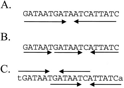
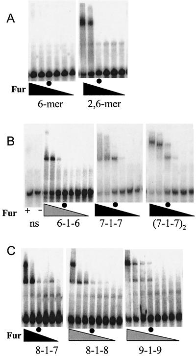
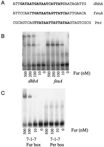
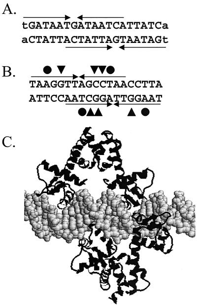
Ferric uptake repressor (Fur) proteins regulate the expression of iron homeostasis genes in response to intracellular iron levels. In general, Fur proteins bind with high affinity to a 19-bp inverted repeat sequence known as the Fur box. An alignment of 19 operator sites recognized by Bacillus subtilis Fur revealed a different conserved 15-bp (7-1-7) inverted repeat present twice within this 19-bp consensus sequence. We demonstrated using electrophoretic mobility shift assays that this 7-1-7 inverted repeat comprises a minimal recognition site for high-affinity binding by Fur. The resulting revised consensus sequence is remarkably similar to a related 7-1-7 inverted repeat sequence recognized by PerR, a Fur paralog. Our analysis of the affinity and stoichiometry of DNA binding by B. subtilis Fur, together with a reinterpretation of previously described studies of Escherichia coli Fur, supports a model in which the 19-bp Fur box represents overlapping recognition sites for two Fur dimers bound to opposite faces of the DNA helix. The resulting recognition complex is reminiscent of that observed for the functionally related protein DtxR. Like Fur, DtxR contains a helix-turn-helix DNA-binding motif, recognizes a 19-bp inverted repeat sequence, and has a typical DNase I footprint of approximately 30 bp. By envisioning a similar mode of DNA recognition for Fur, we can account for the internal symmetries noted previously within the Fur box, the tendency of Fur to extend into adjacent regions of DNA in a sequence-selective manner, and the observed patterns of DNA protection against enzymatic and chemical probes.


Figures

FIG. 1.
FIG. 1.
Comparison of models to explain the Fur box consensus sequence. (A) The Fur box is classically defined as a 19-bp inverted repeat sequence, originally envisioned to bind a single Fur dimer. (B) An alternative view proposes that Fur binds to repeated arrays of three or more copies of the hexamer GATAAT (13, 15). According to this model, the classic Fur box is three GATAAT motifs in a head-to-head-to-tail (6-6-1-6) array. (C) We propose that the 19-bp Fur box results from two overlapping heptamer inverted repeats [(7-1-7)2] that together define a 21-bp sequence.
FIG. 2.
FIG. 2.
Binding of B. subtilis Fur to model oligonucleotide substrates. The final concentrations of Fur protein (monomer) in the reaction mixtures were 0, 1, 10, 100, 500, and 1,000 nM in the panels with six lanes (solid triangles) and 0, 10, 50, 75, 100, 200, 500, and 1,000 nM in the panels with eight lanes (shaded triangles). For ease of comparison between experiments, the lanes containing 100 nM Fur (monomer) are indicated by circles. (A) Binding of Fur protein to oligonucleotides containing either one or two copies of the GATAAT hexamer motif. (B) Comparison of Fur protein binding to the 6-1-6, 7-1-7, and (7-1-7)2 substrates. A nonspecific DNA fragment (ns) (see Table 2) was included as a control with either 1 μM Fur (lane +) or no added Fur (lane −). (C) Effect of additional flanking bases on formation of the lower-mobility complex (see Table 2 for a summary). Note that these studies were not done in parallel with those whose results are shown in panel B, so the absolute affinities cannot be compared.
FIG. 3.
FIG. 3.
Determination of the stoichiometry of Fur-DNA complexes by native PAGE. (A) Logarithms of the relative mobilities of Fur-DNA and marker proteins (versus the mobility of bromophenol blue) as a function of polyacrylamide concentration. The complexes used were Fur-(7-1-7) (□) and Fur-[(7-1-7)2] (○). The marker proteins used were carbonic anhydrase (⧫), α-lactalbumin (▪), bovine serum albumin dimer (•), bovine serum albumin monomer (▾), and ovalbumin (▴). (B) Determination of the apparent molecular weights of the Fur-(7-1-7) and Fur-[(7-1-7)2] complexes. The negative slopes of the mobility lines in panel A were plotted against the molecular weights of the protein standards (solid symbols), and the apparent masses of the Fur-DNA complexes (open symbols) were determined by interpolation.
FIG. 4.
FIG. 4.
Binding of Fur to naturally occurring operator sites. (A) Sequences of the top strands of the DNA oligonucleotides representing the dhb, feuA, and Per box substrates. (B) Fur binds to the dhbA and feu operators with comparable affinities but forms the lower-mobility complex only with the dhb operator. (C) Fur binds to the 7-1-7 substrate but does not recognize the closely related Per box sequence.
FIG. 5.
FIG. 5.
Comparison of the proposed Fur-DNA complex with the DtxR-DNA complex. (A) Two overlapping 7-1-7 heptamer motifs that generate the classic 19-bp Fur box binding site. (B) Two overlapping imperfect inverted repeats that generate the 19-bp binding site for DtxR (adapted from reference 7). The critical contacts for protein-DNA recognition include the interaction of DtxR Gln43 (triangles) with the G·C base pairs indicated and the interaction of a thymine methyl group with the Ser37-Pro39 pair (circles) (adapted from reference 7). (C) Model of the complex formed between DtxR and operator DNA, illustrating the role of two DtxR dimers in recognition.

References

    1. Baichoo, N., T. Wang, R. Ye, and J. D. Helmann. Global analysis of the Bacillus subtilis Fur regulon and the iron starvation stimulon. Mol. Microbiol., in press. - PubMed
    1. Bell, S. D., S. S. Cairns, R. L. Robson, and S. P. Jackson. 1999. Transcriptional regulation of an archaeal operon in vivo and in vitro. Mol. Cell 4:971-982. - PubMed
    1. Bsat, N., L. Chen, and J. D. Helmann. 1996. Mutation of the Bacillus subtilis alkyl hydroperoxide reductase (ahpCF) operon reveals compensatory interactions among hydrogen peroxide stress genes. J. Bacteriol. 178:6579-6586. - PMC - PubMed
    1. Bsat, N., and J. D. Helmann. 1999. Interaction of Bacillus subtilis Fur (ferric uptake repressor) with the dhb operator in vitro and in vivo. J. Bacteriol. 181:4299-4307. - PMC - PubMed
    1. Bsat, N., A. Herbig, L. Casillas-Martinez, P. Setlow, and J. D. Helmann. 1998. Bacillus subtilis contains multiple Fur homologues: identification of the iron uptake (Fur) and peroxide regulon (PerR) repressors. Mol. Microbiol. 29:189-198. - PubMed

Publication types

MeSH terms

Substances