. 2002 Nov;184(21):5935-45.
doi: 10.1128/JB.184.21.5935-5945.2002.

Inducible control of virulence gene expression in Listeria monocytogenes: temporal requirement of listeriolysin O during intracellular infection

Affiliations

Inducible control of virulence gene expression in Listeria monocytogenes: temporal requirement of listeriolysin O during intracellular infection

Christina E Dancz et al. J Bacteriol. 2002 Nov.

Abstract

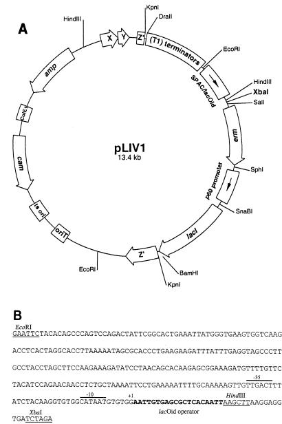
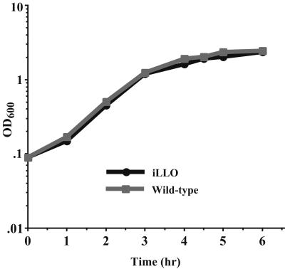
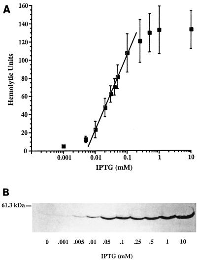
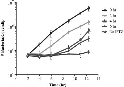
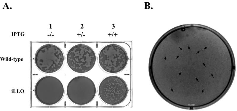
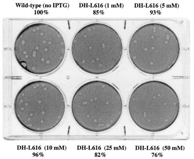
We have constructed a lac repressor/operator-based system to tightly regulate expression of bacterial genes during intracellular infection by Listeria monocytogenes. An L. monocytogenes strain was constructed in which expression of listeriolysin O was placed under the inducible control of an isopropyl-beta-D-thiogalactopyranoside (IPTG)-dependent promoter. Listeriolysin O (LLO) is a pore-forming cytolysin that mediates lysis of L. monocytogenes-containing phagosomes. Using hemolytic-activity assays and Western blot analysis, we demonstrated dose-dependent IPTG induction of LLO during growth in broth culture. Moreover, intracellular growth of the inducible-LLO (iLLO) strain in the macrophage-like cell line J774 was strictly dependent upon IPTG. We have further shown that iLLO bacteria trapped within primary phagocytic vacuoles can be induced to escape into the cytosol following addition of IPTG to the cell culture medium, thus yielding the ability to control bacterial escape from the phagosome and the initiation of intracellular growth. Using the iLLO strain in plaque-forming assays, we demonstrated an additional requirement for LLO in facilitating cell-to-cell spread in L2 fibroblasts, a nonprofessional phagocytic cell line. Furthermore, the efficiency of cell-to-cell spread of iLLO bacteria in L2 cells was IPTG dose dependent. The potential use of this system for determining the temporal requirements of additional virulence determinants of intracellular pathogenesis is discussed.


Figures

FIG. 1.
FIG. 1.
Inducible-expression vector for L. monocytogenes. (A) The pLIV1 vector contains the following sequences: a temperature-sensitive origin of replication (ts ori) and a chloramphenicol resistance gene (cam) for plasmid selection in L. monocytogenes, the ColE1 origin of replication and an ampicillin resistance gene (amp) for cloning and selection in E. coli, an origin of transfer (oriT) to allow conjugal mating of plasmid derivatives from E. coli to L. monocytogenes, a unique XbaI restriction site (shown in bold type) for cloning of genes under the transcriptional control of the SPAC/lacOid IPTG-inducible promoter, tandem copies of the rrnB T1 transcription terminator (T1 terminators) upstream of the SPAC/lacOid region to ensure that transcription of cloned genes initiates only by the SPAC promoter, the L. monocytogenes p60 gene promoter to allow constitutive expression of the lac repressor gene (lacI), and an erythromycin resistance determinant within the expression cassette (erm) for selection of inducible constructs on the chromosome. The inducible gene expression cassette is placed within the L. monocytogenes orfZ gene (Z′), which is immediately followed by a putative transcription terminator and flanked by sufficient DNA to allow homologous recombination. Positions of restriction sites utilized for the construction of pLIV1 are indicated (see Materials and Methods). (B) Nucleotide sequence of the SPAC/lacOid promoter/operator region within pLIV1. The −35 and −10 regions of the SPAC promoter are overlined. The transcription initiation site is indicated as +1. The lacOid operator sequence is shown in bold. Restriction sites are noted and underlined.
FIG. 2.
FIG. 2.
Growth of L. monocytogenes strains in BHI broth. Overnight cultures of iLLO (DP-L3885) or wild-type L. monocytogenes strain 10403S were diluted 1:20 in BHI broth and grown in the presence of 1 mM IPTG for 6 h at 37°C. Culture aliquots were taken every 30 min to 1 h, and the OD600 was determined.
FIG. 3.
FIG. 3.
IPTG induction of LLO is dose dependent. (A) The hemolytic-activity values listed in Table 1 for the iLLO strain (DP-L3885) were plotted versus the IPTG concentration. A best-fit line for linear induction of hemolytic activity is shown. (B) An overnight culture of iLLO strain DP-L3885 was diluted 1:10 in BHI broth into replicate flasks and grown for 5 h at 37°C in the presence of various concentrations of IPTG. The OD600 values of all of the cultures were identical. Culture supernatants were precipitated for 1 h on ice in the presence of 10% (vol/vol) trichloroacetic acid. Samples were centrifuged and resuspended in protein sample buffer. An equivalent amount of each sample was analyzed by sodium dodecyl sulfate-polyacrylamide gel electrophoresis, followed by Western blot assay with a rabbit polyclonal anti-LLO antibody. The position of the LLO bands (58 kDa) relative to that of a molecular size standard (61.3 kDa) is shown.
FIG. 4.
FIG. 4.
Intracellular growth of the iLLO strain in J774 cells. Infection of J774 cells was done as described in Materials and Methods. (A) J774 cells were infected with iLLO (DP-L3885) or LLO-negative (DP-L2161) bacteria in the absence of IPTG. (B) iLLO (DP-L3885) or wild-type (10403S) bacteria were grown for 2.5 h in BHI broth containing 1 mM IPTG prior to infection of J774 cells and maintained in tissue culture medium containing 10 mM IPTG throughout the intracellular growth period.
FIG. 5.
FIG. 5.
Intracellular induction of LLO expression. iLLO (DP-L3885) bacteria were grown in BHI broth in the absence of IPTG and used to infect J774 cell cultures as described in Materials and Methods. Bacteria were added to J774 cells and maintained in medium containing 10 mM IPTG (0 h). Alternatively, bacteria were added to separate J774 cell cultures in the absence of IPTG. At 2-h increments (2, 4, or 6 h) postinfection, culture medium was removed and medium containing 10 mM IPTG was added to J774 cells and maintained throughout the intracellular growth period. One culture received no IPTG addition (No IPTG). Intracellular bacterial counts were determined as described in Materials and Methods.
FIG. 6.
FIG. 6.
LLO induction is required to mediate cell-to-cell spread in L2 fibroblasts. (A) iLLO (DP-L3885) or wild-type (10403S) bacteria were grown in BHI broth without IPTG and added to monolayers of mouse L2 fibroblasts, which were incubated for 1 h. Following washing of infected cell monolayers with PBS, a medium-agarose overlay containing gentamicin was added. Intracellular growth and cell-to-cell spread of bacteria were visualized after 96 h by the formation of clearing zones (plaques) within the L2 monolayers. +/− indicates, respectively, the presence or absence of 1 mM IPTG during the initial 1-h infection period or in the agarose overlay. (B) Magnification (×3) of iLLO plate 2 in panel A. Arrows point to representative pinpoint plaques present within the L2 monolayer.
FIG. 7.
FIG. 7.
Construction of iLLO strain DH-L616. L. monocytogenes strain DH-L616 was constructed by placing the iLLO expression cassette within the L. monocytogenes tRNAArg gene on the chromosome of LLO-negative strain DP-L2161. Plasmid pDH618 was generated by cloning the KpnI fragment harboring the iLLO expression cassette from pLIV1-LLO into pPL2, an L. monocytogenes site-specific phage integration vector (33). Site-specific integration of pDH618 within the tRNAArg gene and verification of the iLLO construct were performed as previously described (33) to yield strain DH-L616.
FIG. 8.
FIG. 8.
IPTG dose-dependent plaque formation in L2 fibroblasts. iLLO (DH-L616) or wild-type (10403S) bacteria were grown in BHI broth without IPTG and added to monolayers of mouse L2 fibroblasts, which were incubated for 1 h. Following washing of the monolayers with PBS, a medium-agarose overlay containing IPTG and gentamicin was added to the monolayers. The concentration of IPTG present during both the initial 1-h infection period and in the agarose overlay is indicated. Cell-to-cell spread of bacteria was visualized after 96 h by the formation of plaques within the monolayers. The average diameter of 9 or 10 plaques per sample was determined and is given as a percentage of that observed for wild-type infection.

References

    1. Aureli, P., G. C. Fiorucci, D. Caroli, G. Marchiaro, O. Novara, L. Leone, and S. Salmaso. 2000. An outbreak of febrile gastroenteritis associated with corn contaminated by Listeria monocytogenes. N. Engl. J. Med. 342:1236-1241. - PubMed
    1. Bayley, H. 1997. Toxin structure: part of a hole? Curr. Biol. 7:R763-R767. - PubMed
    1. Beckwith, J. R., and D. Zisper. 1970. The lactose operon. Cold Spring Harbor Laboratory, Cold Spring Harbor, N.Y.
    1. Biard, D. S. F., M. R. James, A. Cordier, and A. Sarasin. 1992. Regulation of the Escherichia coli lac operon expressed in human cells. Biochim. Biophys. 1130:68-74. - PubMed
    1. Bielecki, J., P. Youngman, P. Connelly, and D. A. Portnoy. 1990. Bacillus subtilis expressing a haemolysin gene from Listeria monocytogenes can grow in mammalian cells. Nature 345:175-176. - PubMed

Publication types

MeSH terms

Associated data