. 2002 Nov;184(21):6026-36.
doi: 10.1128/JB.184.21.6026-6036.2002.

The dilemma of phage taxonomy illustrated by comparative genomics of Sfi21-like Siphoviridae in lactic acid bacteria

Affiliations

The dilemma of phage taxonomy illustrated by comparative genomics of Sfi21-like Siphoviridae in lactic acid bacteria

Caroline Proux et al. J Bacteriol. 2002 Nov.

Abstract

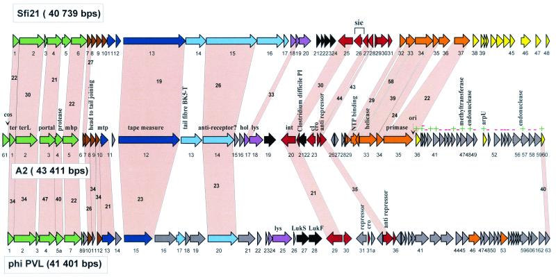
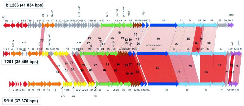
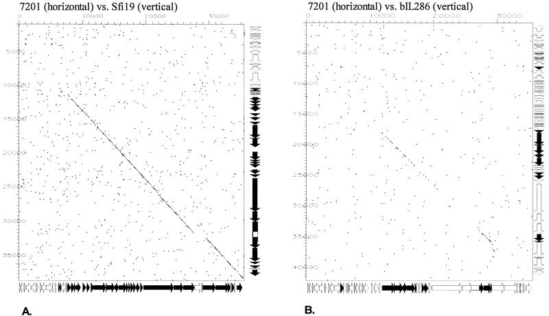
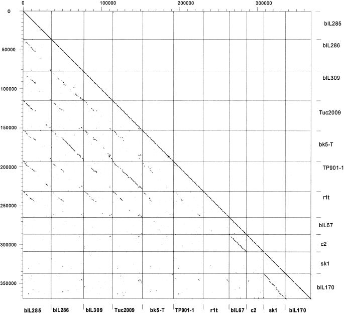
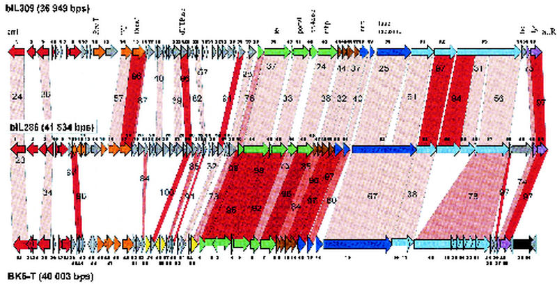
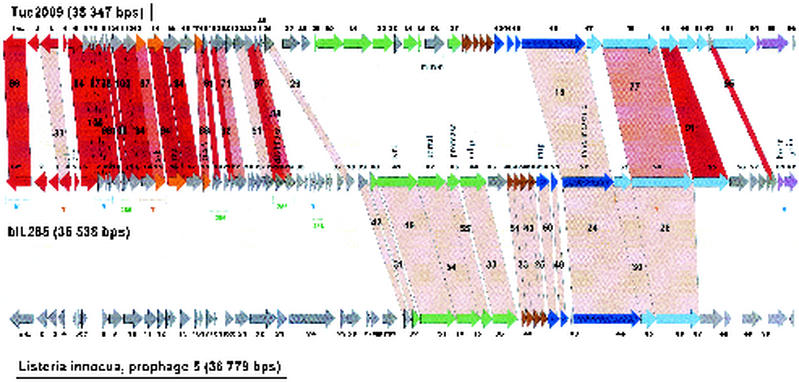
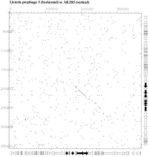
The complete genome sequences of two dairy phages, Streptococcus thermophilus phage 7201 and Lactobacillus casei phage A2, are reported. Comparative genomics reveals that both phages are members of the recently proposed Sfi21-like genus of Siphoviridae, a widely distributed phage type in low-GC-content gram-positive bacteria. Graded relatedness, the hallmark of evolving biological systems, was observed when different Sfi21-like phages were compared. Across the structural module, the graded relatedness was represented by a high level of DNA sequence similarity or protein sequence similarity, or a shared gene map in the absence of sequence relatedness. This varying range of relatedness was found within Sfi21-like phages from a single species as demonstrated by the different prophages harbored by Lactococcus lactis strain IL1403. A systematic dot plot analysis with 11 complete L. lactis phage genome sequences revealed a clear separation of all temperate phages from two classes of virulent phages. The temperate lactococcal phages share DNA sequence homology in a patchwise fashion over the nonstructural gene cluster. With respect to structural genes, four DNA homology groups could be defined within temperate L. lactis phages. Closely related structural modules for all four DNA homology groups were detected in phages from Streptococcus or Listeria, suggesting that they represent distinct evolutionary lineages that have not uniquely evolved in L. lactis. It seems reasonable to base phage taxonomy on data from comparative genomics. However, the peculiar modular nature of phage evolution creates ambiguities in the definition of phage taxa by comparative genomics. For example, depending on the module on which the classification is based, temperate lactococcal phages can be classified as a single phage species, as four distinct phage species, or as two if not three different phage genera. We propose to base phage taxonomy on comparative genomics of a single structural gene module (head or tail genes). This partially phylogeny-based taxonomical system still mirrors some aspects of the current International Committee on Taxonomy in Virology classification system. In this system the currently sequenced lactococcal phages would be grouped into five genera: c2-, sk1, Sfi11-, r1t-, and Sfi21-like phages.


Figures

FIG. 1.
FIG. 1.
Alignment of the genetic maps from the temperate cos site S. thermophilus phage Sfi21, L. casei phage A2, and S. aureus phage PVL, all members of the proposed Sfi21-like genus of Siphoviridae. The ORFs are color coded according to their predicted function deduced from a set of criteria consisting of either their database matches, the synteny argument with phage lambda (18), protein analysis (38), transcription analysis (55), or a mixture of these criteria. Green, predicted DNA packaging and head morphogenesis genes; brown, predicted head-to-tail joining genes; dark blue, tail morphogenesis genes; light blue, tail fiber genes; mauve, lysis genes; black, predicted lysogeny conversion genes; red, lysogeny genes; orange, DNA replication genes; yellow, transcription regulation genes (this region is relatively undefined in A2: ORFs with database match are denoted with a green +, and those lacking a match are denoted with a red −). Selected genes from the A2 phage genome are denoted. Genes encoding proteins that showed significant amino acid sequence similarity are linked by grey shading, and the percentage of amino acid identity is indicated. The phage genomes are depicted following the convention of placing the DNA packaging genes at the left end.
FIG. 2.
FIG. 2.
Alignment of the genetic maps from the L. lactis prophage bIL286 with the virulent S. thermophilus phages 7201 and SfiI9. The open reading frames are color coded according to their predicted function as in Fig. 1. Selected genes or genomic features are denoted. Genes encoding proteins that showed significant amino acid sequence similarity are linked by red shading, and the percentage of amino acid identity is indicated. The degree of amino acid identity (>90, >80, >70, and <70%) is reflected in the color intensity of the red shading. Note that the genomes of the virulent phages were rearranged to allow an easier comparison with the prophage bIL286. The natural ends of the streptococcal phage DNA flank the indicated cos sites.
FIG. 3.
FIG. 3.
Dot plot analysis for the S. thermophilus phage 7201 genome versus the S. thermophilus phage Sfi19 genome (A) and the S. thermophilus phage 7201 genome versus the L. lactis prophage bIL286 genome (B). The comparison window was 50 bp, and the stringency was 30 bp. Genes sharing nucleotide sequence similarity are highlighted in black on the gene maps (see Fig. 2 for an expanded view of the maps) of the phages depicted at the sides of the dot plot.
FIG. 4.
FIG. 4.
Dot plot matrix calculated for the complete genomes of the L. lactis prophages bIL285, bIL286, and bIL309, all from the genome sequence of L. lactis strain IL1403 (13), of the temperate L. lactis phages Tuc2009 (P335 lactococcal species, proposed Sfi11-like genus of pac site Siphoviridae), BK5-T (BK5-T lactococcal species, proposed Sfi21-like genus of cos site Siphoviridae), TP901-1 (P335 lactococcal species, proposed Sfi11-like genus of pac site Siphoviridae), r1t (P335 lactococcal species, new r1t/LC3-like genus (?) of cos site Siphoviridae) and of the virulent L. lactis phages bIL67 and c2 (both c2 lactococcal species, c2-like genus of Siphoviridae) and sk1 and bIL170 (936 species of lactococcal phages, new sk1-like genus (?) of Siphoviridae). The prophages and temperate phages are aligned so that the phage integrase is at the utmost left (top) of each indicated square, followed by the lysogeny and DNA replication modules; in the center are the structural genes (DNA packaging, head, and tail genes); at the right (bottom) of each indicated square are tail fiber and lysis genes. The virulent phages are given with the late structural genes at the right and the early nonstructural genes at the left. The BK5-T and c2 sequences were rearranged or inversed, respectively, with respect to the database entry. The left y axis provides a scale in kilobases; the right y axis and the bottom x axis identify the phage genomes that were compared in the corresponding square. The dot matrix was calculated using Dotter (50). The comparison window was 50 bp, and the stringency was 30 bp.
FIG. 5.
FIG. 5.
Alignment of the genetic maps from the L. lactis prophage bIL309 with the L. lactis prophage bIL286 and temperate L. lactis phage BK5-T. The open reading frames are color coded according to their predicted function as in Fig. 1. Selected genes or genomic features are denoted. Genes encoding proteins that showed significant amino acid sequence are linked by red shading, and the percentage of amino acid identity is indicated. The degree of amino acid identity (>90, >80, >70, or <70%) is reflected in the color intensity of the red shading.
FIG. 6.
FIG. 6.
Alignment of the genetic maps from the temperate L. lactis phage Tuc2009 with L. lactis prophage bIL285 and L. innocua prophage 5. The open reading frames are color coded according to their predicted function as in Fig. 1. Selected genes from bIL285 are annotated. Genes encoding proteins that showed significant amino acid sequence similarity are linked by red shading, and the percentage of amino acid identity is indicated. The degree of amino acid identity (>90, >80, >70, or <70%) is reflected in the color intensity of the red shading. The brackets under the bIL285 gene map demarcate the patchwise sequence sharing of bIL285 genes with other lactococcal phages (B, BK5-T, T, TP901-1; 286, bIL286), possibly identifying genes whose proteins have to interact in a common function (see the text).
FIG. 7.
FIG. 7.
Dot plot analysis for the L. innocua prophage 5 (29) genome versus the L. lactis prophage bIL285 genome. The comparison window was 50 bp, and the stringency was 30 bp. Genes sharing nucleotide sequence similarity are highlighted in black on the gene maps (see Fig. 6 for an expanded view of the prophage maps) of the phages depicted at the sides of the dot plot.

References

    1. Ackermann, H.-W. 1999. Tailed bacteriophages: the order Caudovirales. Adv. Virus Res. 51:135-201. - PMC - PubMed
    1. Allison, G. E., D. Angeles, N. Tran-Dinh, and N. Verma. 2002. Complete genomic sequence of SfV, a serotype-converting temperate bacteriophage of Shigella flexneri. J. Bacteriol. 184:1974-1987. - PMC - PubMed
    1. Altschul, S. F., T. L. Madden, A. A. Schaffer, J. Zhang, Z. Zhang, W. Miller, and D. J. Lipman. 1997. Gapped BLAST and PSI-BLAST: a new generation of protein database search programs. Nucleic Acids Res. 25:3389-3402. - PMC - PubMed
    1. Alvarez, M. A., M. Herrero, and J. E. Suarez. 1998. The site-specific recombination of the Lactobacillus species bacteriophage A2 integrates in Gram-positive and Gram-negative bacteria. Virology 250:185-193. - PubMed
    1. Botstein, D. 1980. A theory of modular evolution for bacteriophages. Ann. N. Y. Acad. Sci. 354:484-491. - PubMed

Publication types

Associated data