. 2002 Oct 15;99(21):13653-8.
doi: 10.1073/pnas.222538099. Epub 2002 Oct 8.

SIRT3, a human SIR2 homologue, is an NAD-dependent deacetylase localized to mitochondria

Affiliations

SIRT3, a human SIR2 homologue, is an NAD-dependent deacetylase localized to mitochondria

Patrick Onyango et al. Proc Natl Acad Sci U S A. .

Abstract

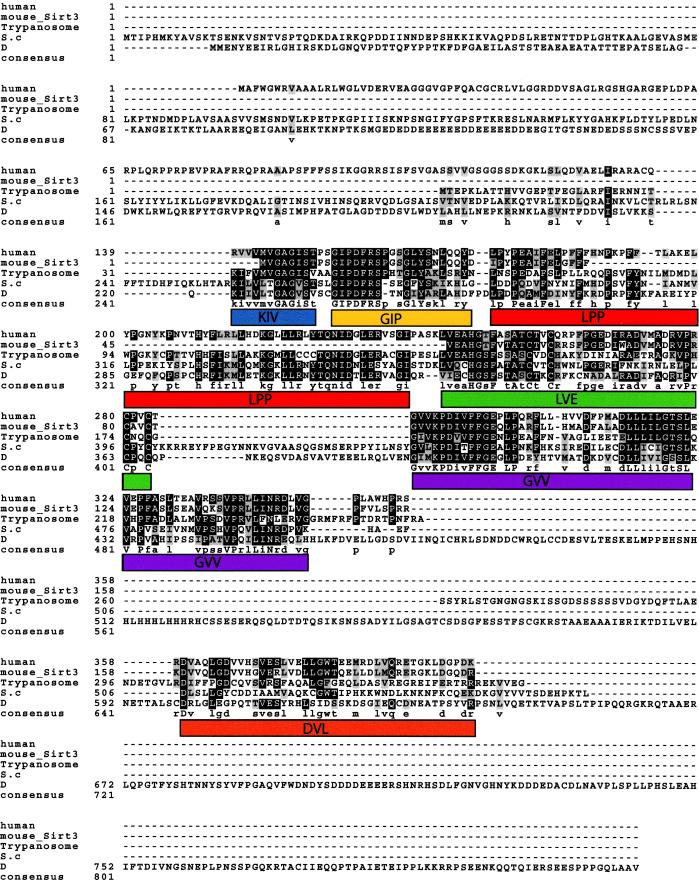
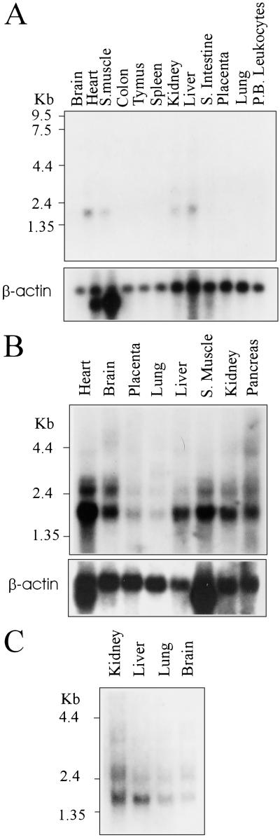
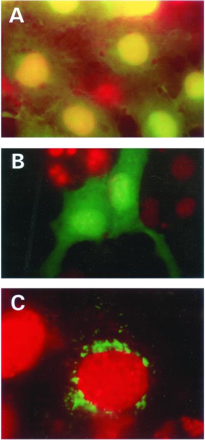
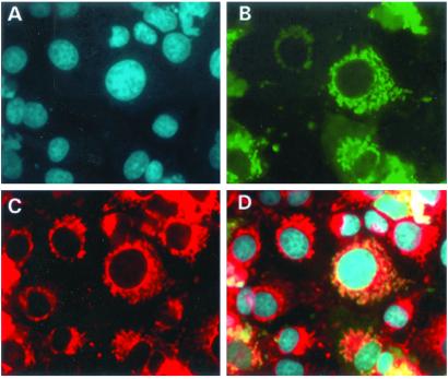
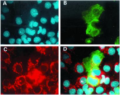
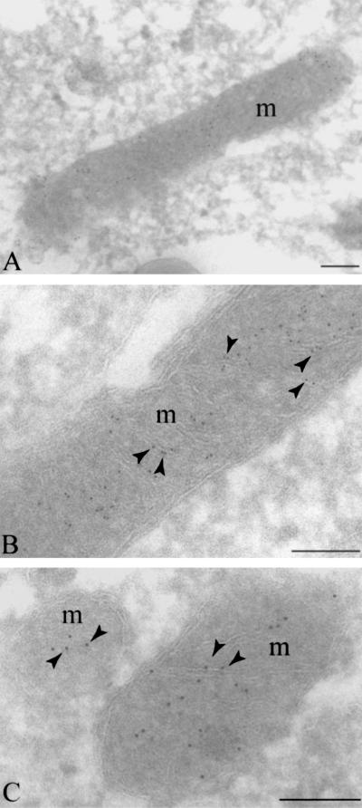
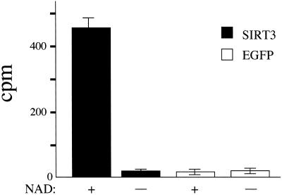
The SIR2 (silent information regulator 2) gene family has diverse functions in yeast including gene silencing, DNA repair, cell-cycle progression, and chromosome fidelity in meiosis and aging. Human homologues, termed sirtuins, are highly conserved but are of unknown function. We previously identified a large imprinted gene domain on 11p15.5 and investigated the 11p15.5 sirtuin SIRT3. Although this gene was not imprinted, we found that it is localized to mitochondria, with a mitochondrial targeting signal within a unique N-terminal peptide sequence. The encoded protein was found also to possess NAD(+)-dependent histone deacetylase activity. These results suggest a previously unrecognized organelle for sirtuin function and that the role of SIRT3 in mitochondria involves protein deacetylation.


Figures

Figure 1
Figure 1
Multiple sequence alignment of the mouse and human SIRT3 and trypanosoma, yeast (S.c), and Drosophila (D) SIR2 proteins. Identities are shaded black, and conserved substitutions are shown in gray. The cutoff for inclusion in the consensus is a 50% match in all the aligned proteins at a given position. Note the relative loss in conservation at the first 142 aa of SIRT3. The overall similarity between the mouse and the human gene is the best, understandably because of a more recent divergence in evolution, compared with the other organisms included in this analysis. Deduced motifs are represented by colored boxes and are given names derived from the first three letters shared from the consensus in all the proteins or, if conserved, in at least three of the five sequences. The motifs are: KIV, blue; GIP, yellow; LPP, red; LVE, green; GVV, purple; and DVL, orange. The KIV and GIP motifs span the conserved Sir2p core domain previously shown to be important in silencing (27).
Figure 2
Figure 2
Expression profile of SIRT3 on a panel of multiple human tissue Northern blot. (A) A 2.1-kb SIRT3 transcript detectable in the heart, skeletal muscle, kidney, and liver. β-actin probe is included as a loading control. (B) Expression of SIRT3 is detectable in all the tissues tested, although only a low level of expression can be seen is the placenta and lung. (Lower) β-actin was used again as a loading control. (C) Expression analysis of SIRT3 in fetal tissues. From loading controls not included, we deduced that the expression level in fetal tissues was equal in all the tested tissues.
Figure 3
Figure 3
Cellular localization of SIRT3 full-length and truncated proteins in Cos7 cells. (A) Shown are cells transfected with vector plasmid. Strong expression of the EGFP in the nucleus and also expression in the cytoplasm is evident. Nuclei were stained with DAPI and colored red, hence the yellow coloration when the green and red signals are superimposed in the nucleus. (B) The N-terminal (1–142 aa) deleted SIR2T3 gene fused in-frame to the EGFP reading frame was transfected into these cells. Note the diffused expression of the chimeric EGFP protein throughout the cell. Unlike with the EGFP shown above, there was no preferential expression of the chimeric protein in the nucleus. Nuclei were detected by counterstaining as described for A. (C) SIR2T3 fused to EGFP (green), localized predominantly around the nuclear periphery. Red shows the nucleus detected by counterstaining with DAPI .
Figure 4
Figure 4
Subcellular localization of the full-length SIRT3 gene in Cos7 cells. A shows DAPI staining of the nuclei, and B depicts the localization of the SIRT3-EGFP protein. Shown in red in C is the mitochondrial marker, Mitotracker. D shows superimposed images of AC. It is clear from D that SIRT3 localizes to the mitochondria.
Figure 5
Figure 5
Subcellular localization of truncated SIRT3 in Cos7 cells. (A) DAPI staining of cell nuclei (blue). (B) SIRT3–EGFP fusion construct transfected into the cells. Green shows the localization of the protein predominantly throughout the cytoplasm. (C) Cells stained with the mitochondrial marker, Mitotracker (red). (D) Superimposition of AC. Note that the fusion construct does not colocalize with the mitochondrial marker.
Figure 6
Figure 6
Immunoelectron microscopy showing localization of SIRT3–EGFP to the mitochondrial cristae. (A) Indirect immunogold labeling of ultrathin cryosections of human cervical carcinoma (SiHa) cells demonstrates SIRT3–EGFP label (5 nm gold) in plain association with mitochondria in three independent images (AC). High-magnification images reveal label clearly associated with mitochondrial cristae (B and C). m, mitochondria; arrowheads, cristae labeling. (Bars, 0.5 μm.)
Figure 7
Figure 7
Protein deacetylase activity of purified SIRT3. Assay for histone deacetylase activity in SIR2T3 and in plasmid vector expressing EGFP only in the presence and absence of 200 mM NAD+. The histone deacetylase activity (cpm) is plotted on the y axis. Experiments were performed in triplicate.

References

    1. Kuo M H, Allis C D. BioEssays. 1998;20:615–626. - PubMed
    1. Aparicio O M, Billington B L, Gottschling D E. Cell. 1991;66:1279–1287. - PubMed
    1. Dutnall R N, Pillus L. Cell. 2001;105:161–164. - PubMed
    1. Smith J S, Boeke J D. Genes Dev. 1997;11:241–254. - PubMed
    1. Kaeberlein M, McVey M, Guarente L. Genes Dev. 1999;13:2570–2580. - PMC - PubMed

Publication types

MeSH terms