. 2002 Oct 15;99(21):13729-34.
doi: 10.1073/pnas.212515299. Epub 2002 Oct 8.

Soluble peptide-MHC monomers cause activation of CD8+ T cells through transfer of the peptide to T cell MHC molecules

Affiliations

Soluble peptide-MHC monomers cause activation of CD8+ T cells through transfer of the peptide to T cell MHC molecules

Qing Ge et al. Proc Natl Acad Sci U S A. .

Abstract

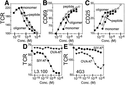
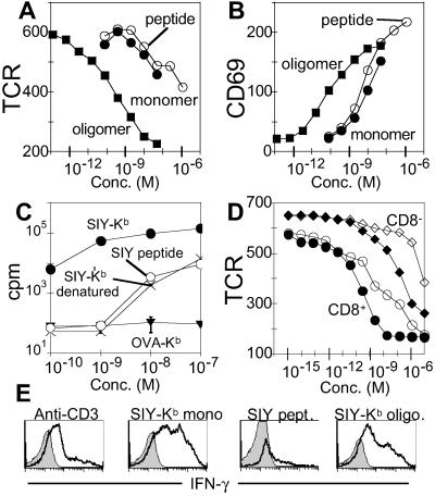
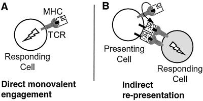
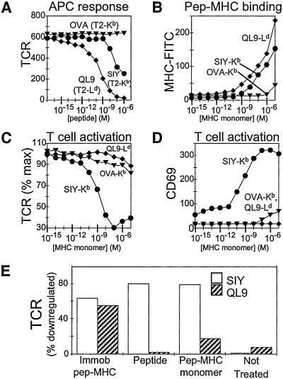
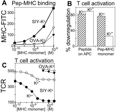
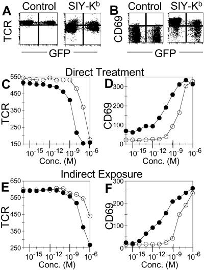
T cell receptor (TCR)-mediated activation of CD4(+) T cells is known to require multivalent engagement of the TCR by, for example, oligomeric peptide-MHC complexes. In contrast, for CD8(+) T cells, there is evidence for TCR-mediated activation by univalent engagement of the TCR. We have here compared oligomeric and monomeric L(d) and K(b) peptide-MHC complexes and free peptide as stimulators of CD8(+) T cells expressing the 2C TCR. We found that the monomers are indeed effective in activating naive and effector CD8(+) T cells, but through an unexpected mechanism that involves transfer of peptide from soluble monomers to T cell endogenous MHC (K(b)) molecules. The result is that T cells, acting as antigen-presenting cells, are able to activate other naive T cells.


Figures

Figure 1
Figure 1
Activation of CD8+ T cell clones by class I MHC monomers and oligomers. (A–C) Response of a T cell clone expressing the 2C TCR (L3.100) to MHC monomers, oligomers, and free peptide. (A) TCR down-regulation in response to 3-h incubation with SIY-Kb oligomer (filled squares), SIY-Kb monomer (filled circles), and SIY peptide alone (open circles). Mean fluorescence intensity (MFI) shown on y axis. (B) CD69 up-regulation (MFI) and (C) CD25 up-regulation (MFI) in response to the same treatment as A. (D and E) Specificity of the response. (D) TCR down-regulation (MFI) is observed for L3.100 incubated for 3 h with SIY-Kb monomers (circles) but not for nonspecific OVA-Kb (inverted triangles). (E) Response of a T cell clone specific for OVA-Kb complex (4G3) shows TCR down-regulation (MFI) after 3 h of incubation with OVA-Kb monomers (inverted triangles), but not with SIY-Kb (circles).
Figure 2
Figure 2
Activation of naïve CD8+ T cells by class I MHC monomers and oligomers. (A and B) Response of naïve T cells expressing the 2C TCR to MHC monomers, oligomers, and free peptide. (A) TCR down-regulation in response to 2-h treatment with SIY-Kb oligomer (filled squares), SIY-Kb monomer (filled circles), and SIY peptide alone (open circles). Mean fluorescence intensity (MFI) shown on y axis. (B) CD69 up-regulation (MFI) in response to the same treatment. (C) Proliferation of naïve 2C T cells measured by 3H-thymidine incorporation 3 days after treatment in response to SIY-Kb monomer (filled circles), SIY peptide alone (open circles), denatured (boiled) SIY-Kb monomer (X), and nonspecific OVA-Kb monomer (inverted triangles). (D) TCR down-regulation (MFI) of CD8+ (circles) or CD8 (diamonds) populations present in purified naïve 2C T cells in response to SIY-Kb monomers (filled symbols) or SIY peptide alone (open symbols). (E) Maturation of naïve 2C T-cells in response to MHC monomers, oligomers, and free peptide. Naïve 2C T cells were stimulated with immobilized anti-CD3 antibody (10 μg/ml), SIY-Kb oligomers, SIY-Kb monomers, or free SIY peptide for 3 days. Intracellular IFN-γ production was measured after 6-h restimulation by SIY peptide (bold line). The shaded area corresponds to isotype control staining.
Figure 3
Figure 3
Two potential mechanisms for activation of T cells by MHC monomers. (A) Binding of monomeric peptide–MHC complexes to cell-surface T cell receptors directly leads to activation (direct engagement model). (B) Transfer of peptide from the soluble MHC molecules to endogenous MHC molecules expressed by the T cell indirectly leads to activation of another T cell through interaction with peptide–MHC complexes on the surface of the first cell (representation model). Binding of soluble peptide–MHC to cell-surface TCR (or CD8) could facilitate peptide transfer, although other mechanisms are possible.
Figure 4
Figure 4
CD8+ T cell activation by MHC monomers requires endogenous MHC proteins. 2C T cells expressing Kb, but not Ld, are activated by soluble monomers of the syngeneic SIY-Kb but by not the potent alloantigen QL9-Ld. (A) Response of naïve 2C T cells to 3 h of incubation with peptide-pulsed APCs. T2 cells expressing Kb were pulsed with OVA (inverted triangles) and SIY (circles) peptides, and T2 cells expressing Ld were incubated with QL9 peptide (diamonds). TCR mean fluorescence intensity (MFI) shown on y axis. (B) Binding of fluorescein-labeled SIY-Kb (circles), OVA-Kb (inverted triangles), or QL9-Ld (diamonds) to naïve 2C T cells. Cell-associated fluorescence was measured after incubation with MHC monomers for 30 min at 4°C, washing, and fixation with paraformaldehyde. (C and D) Response of naïve 2C T cells to 3 h incubation with SIY-Kb (circles), OVA-Kb (inverted triangles), or QL9-Ld (diamonds) monomers. (C) TCR down-regulation (percentage of maximum TCR expression), and (D) CD69 up-regulation (MFI). (E) TCR down-regulation of naïve 2C T cells in response to 6-h incubation with plate-bound and soluble monomers or free peptide. Open bars represent SIY or SIY-Kb; shaded bars represent QL9 or QL9-Ld.
Figure 5
Figure 5
Lack of activation of H2-Kb−/−Db−/− T cells by soluble MHC monomers. T cells expressing the 2C TCR, but not H2-Kb/Db, are unable to be efficiently activated by soluble MHC monomers. (A) Binding of fluorescein-labeled MHC monomers to naïve 2C T cells (filled symbols) and 2C+, Kb−/−Db−/− T cells (open symbols). Binding is shown for SIY-Kb (circles) and OVA-Kb (inverted triangles). MFI shown on y axis. (B) Bar graph showing the percent of TCR down-regulation for treatment of naïve 2C T cells (shaded bars) and 2C+, Kb−/−Db−/− T cells (open bars) with peptide-pulsed T2-Kb cells and soluble SIY-Kb monomer. (C) Concentration dependence of TCR down-regulation (MFI) response in Kb+/+Db+/+ (filled symbols) and Kb−/−Db−/− (open symbols) naïve 2C T cells after 3-h incubation with soluble SIY-Kb (circles) and OVA-Kb (inverted triangles).
Figure 6
Figure 6
T cell activation by uptake and representation of antigen. Naïve GFP+ 2C T cells were activated by exposure to GFP 2C T cells that previously had been treated with MHC monomers. (A and B) GFP cells were treated with control media or SIY-Kb for 3 h, washed, and then GFP+ cells were added to the GFP cells in fresh media. The cells were incubated together for an additional 3 h at 37°C. (A) TCR down-regulation and (B) CD69 up-regulation. The horizontal scale is GFP fluorescence level which distinguishes the directly treated (GFP) and indirectly exposed (GFP+) T cells. (C and D) Response of directly treated GFP+ 2C T cells. (C) TCR down-regulation by GFP+ 2C T cells in response to 3-h incubation with SIY-Kb (filled circles) or SIY peptide (open circles). MFI shown on y axis. (D) CD69 up-regulation (MFI) by GFP+ 2C T cells in response to the same treatment. (E and F) Response of indirectly exposed GFP+ 2C T cells. (E) TCR down-regulation (MFI) by GFP+ 2C T cells incubated for 3 h with GFP 2C T cells that had been pretreated with SIY-Kb (filled circles) or SIY peptide (open circles). (F) CD69 up-regulation (MFI) of GFP+ 2C T cells in response to the same stimuli.

References

    1. Roelants G, Forni L, Pernis B. J Exp Med. 1973;137:1060–1077. - PMC - PubMed
    1. Dintzis H M, Dintzis R Z, Vogelstein B. Proc Natl Acad Sci USA. 1976;73:3671–3675. - PMC - PubMed
    1. Metzger H. J Immunol. 1992;149:1477–1487. - PubMed
    1. Abastado J P, Lone Y C, Casrouge A, Boulot G, Kourilsky P. J Exp Med. 1995;182:439–447. - PMC - PubMed
    1. Boniface J J, Rabinowitz J D, Wulfing C, Hampl J, Reich Z, Altman J D, Kantor R M, Beeson C, McConnell H M, Davis M M. Immunity. 1998;9:459–466. - PubMed

Publication types

Substances