. 2002 Oct 15;99(21):13397-402.
doi: 10.1073/pnas.192468299. Epub 2002 Oct 8.

Expanding pyrimidine diphosphosugar libraries via structure-based nucleotidylyltransferase engineering

Affiliations

Expanding pyrimidine diphosphosugar libraries via structure-based nucleotidylyltransferase engineering

William A Barton et al. Proc Natl Acad Sci U S A. .

Abstract

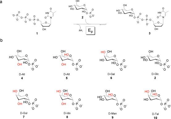
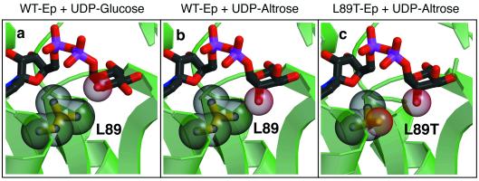
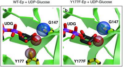
In vitro "glycorandomization" is a chemoenzymatic approach for generating diverse libraries of glycosylated biomolecules based on natural product scaffolds. This technology makes use of engineered variants of specific enzymes affecting metabolite glycosylation, particularly nucleotidylyltransferases and glycosyltransferases. To expand the repertoire of UDP/dTDP sugars readily available for glycorandomization, we now report a structure-based engineering approach to increase the diversity of alpha-d-hexopyranosyl phosphates accepted by Salmonella enterica LT2 alpha-d-glucopyranosyl phosphate thymidylyltransferase (E(p)). This article highlights the design rationale, determined substrate specificity, and structural elucidation of three "designed" mutations, illustrating both the success and unexpected outcomes from this type of approach. In addition, a single amino acid substitution in the substrate-binding pocket (L89T) was found to significantly increase the set of alpha-d-hexopyranosyl phosphates accepted by E(p) to include alpha-d-allo-, alpha-d-altro-, and alpha-d-talopyranosyl phosphate. In aggregate, our results provide valuable blueprints for altering nucleotidylyltransferase specificity by design, which is the first step toward in vitro glycorandomization.


Figures

Fig 1.
Fig 1.
(a) The reaction catalyzed by Ep. (b) Sugar phosphates used for this study. The corresponding deviations from the natural substrate (2) are highlighted in red.
Fig 2.
Fig 2.
Schematic representation of Ep and glucose active-site interactions using the program LIGPLOT (19). The glucose moiety of the product, UDP-glucose, and the residues that interact with it in the Ep active site are shown in ball-and-stick format. Hydrogen bonds are illustrated as dashed lines, and hydrophobic interactions are indicated by half circles.
Fig 3.
Fig 3.
Fitting altrose in the Ep active site. Close-up views of the structure of wild-type Ep and L89T variant bound to UDP-glucose or modeled UDP-altrose. The entire ligand is illustrated in ball-and-stick format, and the C-2 oxygen atom is shown in space-filling representation. Leu-89 and Thr-89 are depicted in both ball-and-stick and space-filling formats. (a) Wild-type (WT) Ep bound to the product, UDP-glucose. Note the space separating the UDP-glucose C-2 oxygen and Leu-89. (b) The activated sugar, UDP-altrose, is shown modeled in the wild-type Ep active site. Note the steric interference generated when attempting to use this C-2 epimer of glucose as a substrate. (c) Modeled in the active site of L89T is the product UDP-altrose. Observe the separation between Thr-89 and the altrose C-2 oxygen.
Fig 4.
Fig 4.
Comparison between wild-type (WT) Ep and Y177F active sites. The ligand, UDP-glucose, is illustrated in ball-and-stick format, as are Gly-147, Tyr-177, and Phe-177. Space-filling representations are shown for the side-chain oxygen in Tyr-177, the main-chain nitrogen of Gly-147, and the C-2 oxygen of UDG. (a) Close-up view of the Ep–UDP-glucose complex active site (left). Note the distance between the backbone nitrogen of Gly-147 and the UDP-glucose C-3 oxygen. An axial oxygen at this position would probably result in steric clashes. (b) A close-up view of the Y177F–UDP-glucose active site. Observe the tilt in the glucose ring in this model. The C-6 oxygen is now in view. Also, notice the slight increase in distance between the nitrogen of Gly-147 and the UDP-glucose C-3 oxygen.
Fig 5.
Fig 5.
Effects of main-chain distortion in the W224H variant. The ligand, UDP-glucose, is illustrated in ball-and-stick format, as are Ep residues Tyr-138, Trp-224, and His-224. (a) The wild-type (WT) Ep active site showing the position of Trp-224 and Tyr-138 relative to the product UDP-glucose. Note the distance between the UDP-glucose C-6 oxygen and the Trp-224 side chain. (b) The W224H active site showing the large movement of His-224 relative to Trp-224 in a. Also, note the large difference in conformation of Tyr-138. It rotates almost 180° to accommodate His-224. Movement of these side chains opens up a large gap surrounding C-6.

References

    1. Thorson J. S., Hosted, T. J., Jr., Jiang, J., Biggins, J. B., Ahlert, J. & Ruppen, M. (2001) Curr. Org. Chem. 5, 139-167.
    1. Solenberg P. J., Matsushima, P., Stack, D. R., Wilkie, S. C., Thompson, R. C. & Baltz, R. H. (1997) Chem. Biol. 4, 195-202. - PubMed
    1. Losey H. C., Peczuh, M. W., Chen, Z., Eggert, U. S., Dong, S. D., Pelczer, I., Kahne, D. & Walsh, C. T. (2001) Biochemistry 40, 4745-4755. - PubMed
    1. Mulichak A. M., Losey, H. C., Walsh, C. T. & Garavito, R. M. (2001) Structure (London) 9, 547-557. - PubMed
    1. Jiang J., Biggins, J. B. & Thorson, J. S. (2001) Angew. Chem. Intl. Ed. Engl. 40, 1502-1505. - PubMed

Publication types

MeSH terms

Substances

LinkOut - more resources