. 2002 Oct 29;99(22):14183-8.
doi: 10.1073/pnas.172517799. Epub 2002 Oct 8.

Cell surface polarization during yeast mating

Affiliations

Cell surface polarization during yeast mating

Michel Bagnat et al. Proc Natl Acad Sci U S A. .

Abstract

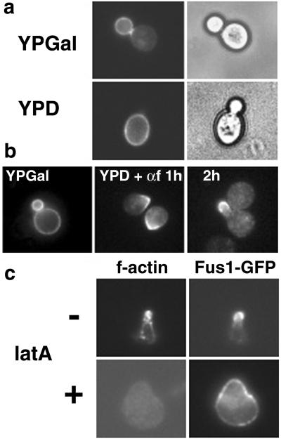
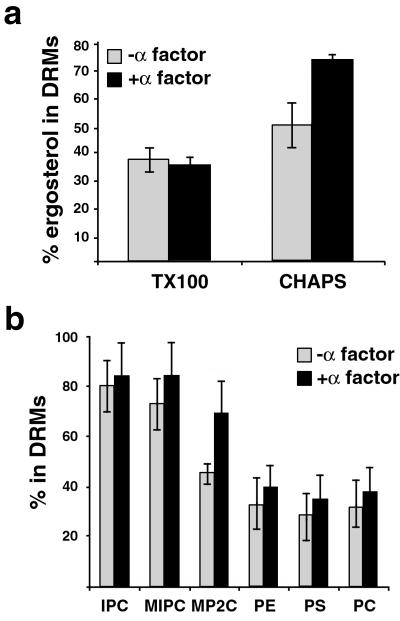
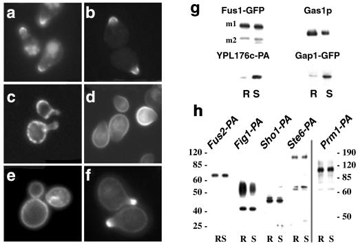
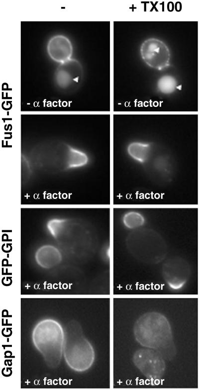
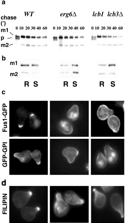
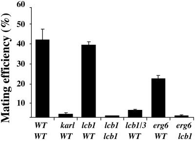
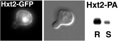
Exposure to mating pheromone in haploid Saccharomyces cerevisiae cells results in the arrest of the cell cycle, expression of mating-specific genes, and polarized growth toward the mating partner. Proteins involved in signaling, polarization, cell adhesion, and fusion are localized to the tip of the mating cell (shmoo) where fusion will eventually occur. The mechanisms ensuring the correct targeting and retention of these proteins are poorly understood. Here we show that in pheromone-treated cells, a reorganization of the plasma membrane involving lipid rafts results in the retention of proteins at the tip of the mating projection, segregated from the rest of the membrane. Sphingolipid and ergosterol biosynthetic mutants fail to polarize proteins to the tip of the shmoo and are deficient in mating. Our results show that membrane microdomain clustering at the mating projection is involved in the generation and maintenance of polarity during mating.


Figures

Fig 1.
Fig 1.
Reorganization of the plasma membrane in α-factor-treated cells. (a) WT cells (RH690-15D) were grown in YPD at 30°C and shifted to YPGal to induce expression Fus1-GFP under the control of the GALS (29) promoter (pMBQ30). Then cells were shifted to YPD for 2 h to turn off Fus1-GFP expression. (b) Fus1-GFP was expressed as in a, and cells were shifted to YPD for 1 h and then treated with α-factor (3 μM) for the indicated times. (c) Actin-dependent localization of Fus1-GFP. Cells were processed as in b and after 2 h of α-factor (3 μM) treatment, and latrunculin A (latA) was added for 1 h more. Cells were fixed and f-actin stained with rhodamine-phalloidin.
Fig 2.
Fig 2.
(a) DRM association of ergosterol in vegetative (−α-factor) and pheromone-treated (+α-factor) cells. The percentage of DRM-associated ergosterol corresponds to ratio of detergent resistant vs. total ergosterol in membranes. The values correspond to the means of three experiments with SD. (b) To determine the pattern of DRM-associated phopholipids, cells were labeled with [32P]orthophosphate with or without the addition of α-factor for 3 h. Cells were lysed, and the fraction of the indicated lipid species in DRM (in CHAPS) was determined as described (23). IPC, inositol-phosphorylceramide; MIPC, mannose-inositol-phosphorylceramide; MP2C, mannose-(inositol phosphorus)2-ceramide; PE, phosphatidylethanolamine; PS, phosphatidylserine; PC, phosphatidylcholine (n = 3). In ref. PS and # (symbol shown in ref. figures) were swapped.
Fig 3.
Fig 3.
Shmoo tip-targeted proteins become DRM-associated. (a) Fus1-GFP localized to the tip of shmoo in WT cells (MBY229) after α-factor (3 μM) induction. A pool of Fus1-GFP localized to the vacuole, probably because of turnover (see Fig. 5). (b) Gas1-derived GFP-GPI (RH690-15D[pMBQ31]). (c) Transferrin receptor-like YPL176c-GFP (RH690-15D). (d) Gap1-GFP (pCK230) in bul1Δ bul2Δ mutant cells (CKY698). Fus1-GFP and GFP-GPI were localized as in WT cells in bul1Δ bul2Δ mutant cells not (not shown). (e) Sterol distribution in control and α-factor polarized cells (f) was visualized after incubation with filipin (9 μg/ml) for 15 min. (g) Cells (MBY229 and CKY698[pCK230]) were preincubated with 5 μM α-factor for 15 min and pulse-labeled with [35S]methionine for 15 min, and after a 45-min chase, the cells were lysed. After extraction with 1% CHAPS and Optiprep density gradient centrifugation, Fus1-GFP and Ga1p-GFP were immunoprecipitated from detergent-resistant (R) and soluble (S) fractions and analyzed by SDS/PAGE and phosphorimaging (m1 and m2 are two mature products of Fus1-GFP). YPL176c-PA (MBY205) and Gas1p (MBY229) DRM-association was analyzed by immunoblotting. Gas1p and Fus1-GFP remained DRM associated as in WT cells in the bul1Δ bul2Δ background (not shown). (h) Fus2, Fig1, Sho1, St6, and Prm1 were tagged with the IgG-binding domain of PA and processed as before and detected by immunoblotting. At least 30% of the protein floated after density gradient centrifugation whereas the bulk of YPL176c-PA remained soluble under the same conditions. The molecular weight is indicated.
Fig 4.
Fig 4.
DRMs are clustered at the tip the mating projections. The distribution of DRM-associated Fus1-GFP was investigated by treating the cells with cold TX100 (1%) for 30 min. In budding cells, Fus1-GFP was partially extractable with cold detergent; the DRM-associated pool was found in patches along the entire surface of the bud. The arrowheads mark the vacuole. In contrast, in α-factor-treated cells Fus1-GFP remained as a continuous detergent-resistant domain at the tip of shmoos. Gap1-GFP, but not GFP-GPI, was sensitive to extraction in α-factor-treated cells.
Fig 5.
Fig 5.
Processing and sorting of shmoo-tip markers in lipid biosynthetic mutants. (a) WT (MBY206), erg6Δ (MBY240), and lcb1–100 lcb3Δ (MBY223) cells were preincubated with 5 μM α-factor for 15 min, pulse-labeled with [35S]methionine for 5 min, and chased for various times at 24°C. Fus1-PA maturation was analyzed by immunoprecipitation, SDS/PAGE, and autoradiography. The position of precursor (p) and to mature products (m1 and m2) is indicated. (b) DRM association of Fus1-PA in WT (MBY206), erg6Δ (MBY240), and lcb1–100 lcb3Δ (MBY223) cells was analyzed as in Fig. 2. (c) Localization of Fus1-GFP (pMBQ30) and GFP-GPI (pMBQ31) in WT (RH690-15D), erg6Δ (RH3622), and lcb1–100 lcb3Δ (RH690-13B) cells. Cells were grown on YPGal and treated with 3 μM α-factor for 3 h. (d) Sterol distribution.
Fig 6.
Fig 6.
Lipid biosynthetic mutants have reduced mating efficiency. Quantitative matings were performed at 24°C for 3 h as described (19). The mating efficiency (diploids/total cells) is given as the means of three experiments with SD. AAY1017 was used as a WT tester. Mating mutant kar1-1 strain (2339) was used as a control.
Fig 7.
Fig 7.
DRM-associated Hxt2 is excluded from the tip of the shmoo. Hxt2-GFP (MBY238) and Hxt2-PA (MBY242) were induced by shifting the cells to low (0.1%) dextrose. Cells were treated with 3 μM α-factor for 3 h and processed as in Fig. 3. Most of Hxt2-PA was found in the DRM fraction (R).

References

    1. van Meer G., Gumbiner, B. & Simons, K. (1986) Nature 322, 639-641. - PubMed
    1. Manes S., Mira, E., Gomez-Mouton, C., Lacalle, R. A., Keller, P., Labrador, J. P. & Martinez, A. C. (1999) EMBO J. 18, 6211-6220. - PMC - PubMed
    1. Bromley S. K., Burack, W. R., Johnson, K. G., Somersalo, K., Sims, T. N., Sumen, C., Davis, M. M., Shaw, A. S., Allen, P. M. & Dustin, M. L. (2001) Annu. Rev. Immunol. 19, 375-396. - PubMed
    1. Brown D. A. & London, E. (1998) Annu. Rev. Cell Dev. Biol. 14, 111-136. - PubMed
    1. Simons K. & Toomre, D. (2000) Nat. Rev. Mol. Cell. Biol. 1, 31-39. - PubMed

MeSH terms

Substances